The healing pattern of a 4 mm proximal infrabony defect was not significantly different from a 2 mm defect adjacent to dental implant in a canine mandible.
Min Kuk An, Hyun Ju Kim, Jin Uk Choi, Kyoung-Hwa Kim, Yong-Moo Lee, In-Chul Rhyu, Yang-Jo Seol
{"title":"The healing pattern of a 4 mm proximal infrabony defect was not significantly different from a 2 mm defect adjacent to dental implant in a canine mandible.","authors":"Min Kuk An, Hyun Ju Kim, Jin Uk Choi, Kyoung-Hwa Kim, Yong-Moo Lee, In-Chul Rhyu, Yang-Jo Seol","doi":"10.5051/jpis.2106420321","DOIUrl":null,"url":null,"abstract":"<p><strong>Purpose: </strong>The purpose of this study was to evaluate and compare the healing patterns of 2-mm and 4-mm proximal infrabony defects adjacent to dental implants in canine mandibles.</p><p><strong>Methods: </strong>Four male beagles were used. Two groups were created: a 2-mm group (n=4) and a 4-mm group (n=4) depending on the horizontal dimension of proximal infrabony defects adjacent to implants. Bone healing patterns between the 2 groups were evaluated and compared at 8 and 16 weeks using radiographic, histological, histomorphometric, and fluorescent labelling analyses.</p><p><strong>Results: </strong>According to microcomputed tomography, the median bone volume fraction, bone mineral density, and the percentage of radiographic distance from the defect bottom to the most coronal bone-to-implant contact (radio-mcBIC) were 32.9%, 0.6 g/cm<sup>3</sup>, and 73.7% (8 weeks) and 45.7%, 0.7 g/cm<sup>3</sup>, and 76.0% (16 weeks) in the 2-mm group and 57.7%, 0.8 g/cm<sup>3</sup>, and 75.7% (8 weeks) and 50.9%, 0.8 g/cm<sup>3</sup>, and 74.7% (16 weeks) in the 4-mm group, respectively. According to histomorphometry, the median bone area fraction, mcBIC and the percentage of BIC amounted to 36.7%, 3.4 mm, and 58.4% (8 weeks) and 49.2%, 3.4 mm, and 70.2% (16 weeks) in the 2-mm group and 50.0%, 3.0 mm, and 64.8% (8 weeks) and 55.7%, 3.0 mm, and 69.6% (16 weeks) in the 4-mm group, respectively. No statistically significant differences were found between the groups for any variables (<i>P</i>>0.05).</p><p><strong>Conclusions: </strong>The proximal defects that measured 2 mm and 4 mm showed similar healing patterns at 8 and 16 weeks, and the top of bone formation in the defects was substantially limited to a maximum of 1.6 mm below the implant shoulder in both groups.</p>","PeriodicalId":48795,"journal":{"name":"Journal of Periodontal and Implant Science","volume":null,"pages":null},"PeriodicalIF":2.2000,"publicationDate":"2022-10-01","publicationTypes":"Journal Article","fieldsOfStudy":null,"isOpenAccess":false,"openAccessPdf":"https://ftp.ncbi.nlm.nih.gov/pub/pmc/oa_pdf/6b/3a/jpis-52-422.PMC9614175.pdf","citationCount":"0","resultStr":null,"platform":"Semanticscholar","paperid":null,"PeriodicalName":"Journal of Periodontal and Implant Science","FirstCategoryId":"3","ListUrlMain":"https://doi.org/10.5051/jpis.2106420321","RegionNum":4,"RegionCategory":"医学","ArticlePicture":[],"TitleCN":null,"AbstractTextCN":null,"PMCID":null,"EPubDate":"","PubModel":"","JCR":"Q2","JCRName":"DENTISTRY, ORAL SURGERY & MEDICINE","Score":null,"Total":0}
引用次数: 0
Abstract
Purpose: The purpose of this study was to evaluate and compare the healing patterns of 2-mm and 4-mm proximal infrabony defects adjacent to dental implants in canine mandibles.
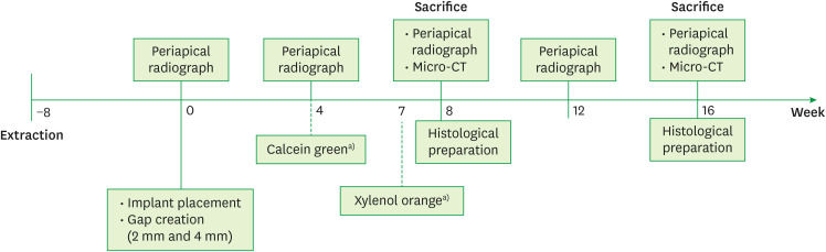
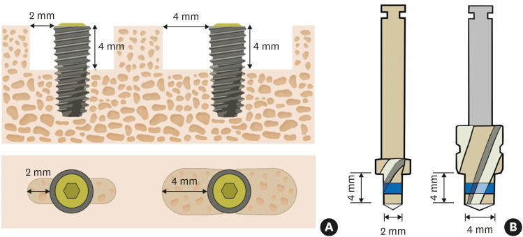
Methods: Four male beagles were used. Two groups were created: a 2-mm group (n=4) and a 4-mm group (n=4) depending on the horizontal dimension of proximal infrabony defects adjacent to implants. Bone healing patterns between the 2 groups were evaluated and compared at 8 and 16 weeks using radiographic, histological, histomorphometric, and fluorescent labelling analyses.
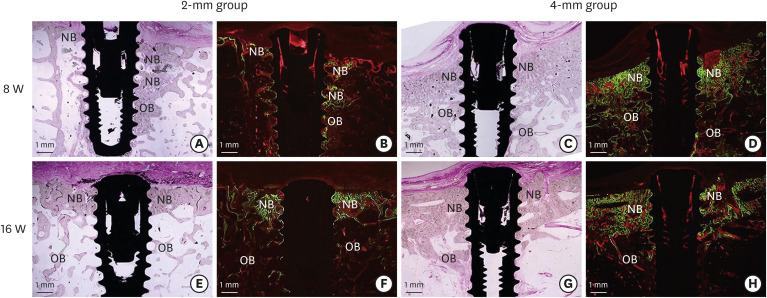
Results: According to microcomputed tomography, the median bone volume fraction, bone mineral density, and the percentage of radiographic distance from the defect bottom to the most coronal bone-to-implant contact (radio-mcBIC) were 32.9%, 0.6 g/cm3, and 73.7% (8 weeks) and 45.7%, 0.7 g/cm3, and 76.0% (16 weeks) in the 2-mm group and 57.7%, 0.8 g/cm3, and 75.7% (8 weeks) and 50.9%, 0.8 g/cm3, and 74.7% (16 weeks) in the 4-mm group, respectively. According to histomorphometry, the median bone area fraction, mcBIC and the percentage of BIC amounted to 36.7%, 3.4 mm, and 58.4% (8 weeks) and 49.2%, 3.4 mm, and 70.2% (16 weeks) in the 2-mm group and 50.0%, 3.0 mm, and 64.8% (8 weeks) and 55.7%, 3.0 mm, and 69.6% (16 weeks) in the 4-mm group, respectively. No statistically significant differences were found between the groups for any variables (P>0.05).
Conclusions: The proximal defects that measured 2 mm and 4 mm showed similar healing patterns at 8 and 16 weeks, and the top of bone formation in the defects was substantially limited to a maximum of 1.6 mm below the implant shoulder in both groups.
期刊介绍:
Journal of Periodontal & Implant Science (JPIS) is a peer-reviewed and open-access journal providing up-to-date information relevant to professionalism of periodontology and dental implantology. JPIS is dedicated to global and extensive publication which includes evidence-based original articles, and fundamental reviews in order to cover a variety of interests in the field of periodontal as well as implant science.
 求助内容:
求助内容: 应助结果提醒方式:


