{"title":"Microvascular Changes in the Cystic Lesion of Branch Retinal Vein Occlusion Imaged by Swept-Source Optical Coherence Tomography Angiography.","authors":"Satoko Araki, Susumu Sakimoto, Daiki Shiozaki, Chihiro Ueda, Chikako Hara, Yoko Fukushima, Kaori Sayanagi, Hirokazu Sakaguchi, Kohji Nishida","doi":"10.1159/000525497","DOIUrl":null,"url":null,"abstract":"<p><strong>Introduction: </strong>This study aimed to describe the quantitative features of the microvasculature in the cystic lesions of branch retinal vein occlusion (BRVO).</p><p><strong>Methods: </strong>A total of 43 eyes with BRVO, treated with anti-vascular endothelial growth factor therapy, were analyzed. Using wide-field swept-source optical coherence tomography angiography (OCTA), en face OCT images were obtained by depth-integrated reflectivity of the retina, and vascular density (VD), vascular length (VL), vascular lacunarity, and fractal dimension (FD) were evaluated in a 12 × 12-mm area of retinal nonperfusion.</p><p><strong>Results: </strong>The mean area of affected lesions was 38.7 ± 19.8 mm<sup>2</sup>, and cystic lesions were 8.5 ± 10.1 mm<sup>2</sup>. VD, VL, and FD were significantly decreased in the cystic lesions compared to other affected lesions in the same eyes (<i>p</i> = 0.0010, <i>p</i> = 0.0001, and <i>p</i> = 0.0003, respectively) and in all eyes (<i>p</i> = 0.0281, <i>p</i> = 0.0050, and <i>p</i> < 0.0001, respectively). VD in cystic lesions within the vascular arcade (25 eyes) correlated with best-corrected visual acuity on OCTA (<i>r</i> = -0.433, and <i>p</i> = 0.0492).</p><p><strong>Conclusions: </strong>Vascular structure in the cystic lesions was unpreserved compared to the other lesions in BRVO. These findings may help in understanding the pathophysiology of retinal edema in BRVO.</p>","PeriodicalId":9075,"journal":{"name":"Biomedicine Hub","volume":"7 2","pages":"99-105"},"PeriodicalIF":0.0000,"publicationDate":"2022-08-16","publicationTypes":"Journal Article","fieldsOfStudy":null,"isOpenAccess":false,"openAccessPdf":"https://ftp.ncbi.nlm.nih.gov/pub/pmc/oa_pdf/23/ce/bmh-0007-0099.PMC9574207.pdf","citationCount":"1","resultStr":null,"platform":"Semanticscholar","paperid":null,"PeriodicalName":"Biomedicine Hub","FirstCategoryId":"1085","ListUrlMain":"https://doi.org/10.1159/000525497","RegionNum":0,"RegionCategory":null,"ArticlePicture":[],"TitleCN":null,"AbstractTextCN":null,"PMCID":null,"EPubDate":"2022/5/1 0:00:00","PubModel":"eCollection","JCR":"","JCRName":"","Score":null,"Total":0}
引用次数: 1
Abstract
Introduction: This study aimed to describe the quantitative features of the microvasculature in the cystic lesions of branch retinal vein occlusion (BRVO).
Methods: A total of 43 eyes with BRVO, treated with anti-vascular endothelial growth factor therapy, were analyzed. Using wide-field swept-source optical coherence tomography angiography (OCTA), en face OCT images were obtained by depth-integrated reflectivity of the retina, and vascular density (VD), vascular length (VL), vascular lacunarity, and fractal dimension (FD) were evaluated in a 12 × 12-mm area of retinal nonperfusion.
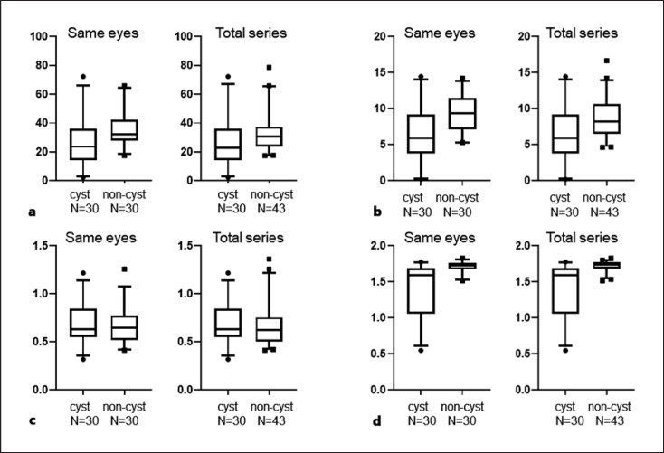
Results: The mean area of affected lesions was 38.7 ± 19.8 mm2, and cystic lesions were 8.5 ± 10.1 mm2. VD, VL, and FD were significantly decreased in the cystic lesions compared to other affected lesions in the same eyes (p = 0.0010, p = 0.0001, and p = 0.0003, respectively) and in all eyes (p = 0.0281, p = 0.0050, and p < 0.0001, respectively). VD in cystic lesions within the vascular arcade (25 eyes) correlated with best-corrected visual acuity on OCTA (r = -0.433, and p = 0.0492).
Conclusions: Vascular structure in the cystic lesions was unpreserved compared to the other lesions in BRVO. These findings may help in understanding the pathophysiology of retinal edema in BRVO.


 求助内容:
求助内容: 应助结果提醒方式:


