Christy L Pylypjuk, Chelsea Day, Yasmine ElSalakawy, Gregory J Reid
{"title":"The Significance of Exposure to Pregestational Type 2 Diabetes in Utero on Fetal Renal Size and Subcutaneous Fat Thickness.","authors":"Christy L Pylypjuk, Chelsea Day, Yasmine ElSalakawy, Gregory J Reid","doi":"10.1155/2022/3573963","DOIUrl":null,"url":null,"abstract":"<p><strong>Objectives: </strong>To determine the relationship between exposure to pregestational type 2 diabetes (T2D) and renal size and subcutaneous fat thickness in fetuses during routine obstetrical ultrasound.</p><p><strong>Methods: </strong>This was a case-control study (January 1, 2019 to December 31, 2019). Routine obstetrical ultrasounds performed between 18 and 22 weeks' gestation at a tertiary-care fetal assessment unit were reviewed. \"Cases\" comprised ultrasounds of fetuses exposed to pregestational T2D in utero. The control group was assembled from ultrasounds of healthy controls. Postprocessing measurements of fetal renal size and abdominal wall thickness from stored images were performed by two independent observers, and findings were compared between groups.</p><p><strong>Results: </strong>There were 54 cases and 428 ultrasounds of healthy controls. The mean maternal age of cases was 32.1 years (SD 6.2) compared to 33.2 years (SD 5.3) for healthy controls, and the majority of ultrasounds were performed in multiparous patients (83%). At the 18 to 22 week ultrasound, there was a significant reduction in renal size amongst fetuses exposed to maternal T2D in utero compared to controls; among cases, the mean renal width was 8.0 mm (95% CI 7.8-8.1) compared to 11.4 mm (95% CI 10.6-12.7) in controls (<i>p</i> < 0.0001); the mean renal thickness among cases was 8.1 mm (95% CI 7.9-8.2) compared to 11.5 mm (95% CI 10.7-12.9) in controls (<i>p</i>=0.001). There was no obvious difference in estimated fetal weight between groups, yet fetuses exposed to maternal T2D had increased subcutaneous abdominal wall fat thickness at this early gestational age (<i>p</i>=0.008).</p><p><strong>Conclusions: </strong>Fetal renal size in cases exposed to pregestational T2D is significantly smaller compared to controls, and subcutaneous abdominal wall fat is significantly thicker. Given emerging evidence about the developmental origins of disease, further study is needed to correlate the association between fetal renal size and fat distribution in the fetus and the long-term risk of chronic renal disease and diabetes in these offspring.</p>","PeriodicalId":14177,"journal":{"name":"International Journal of Nephrology","volume":" ","pages":"3573963"},"PeriodicalIF":1.7000,"publicationDate":"2022-06-30","publicationTypes":"Journal Article","fieldsOfStudy":null,"isOpenAccess":false,"openAccessPdf":"https://www.ncbi.nlm.nih.gov/pmc/articles/PMC9262542/pdf/","citationCount":"2","resultStr":null,"platform":"Semanticscholar","paperid":null,"PeriodicalName":"International Journal of Nephrology","FirstCategoryId":"1085","ListUrlMain":"https://doi.org/10.1155/2022/3573963","RegionNum":0,"RegionCategory":null,"ArticlePicture":[],"TitleCN":null,"AbstractTextCN":null,"PMCID":null,"EPubDate":"2022/1/1 0:00:00","PubModel":"eCollection","JCR":"Q3","JCRName":"UROLOGY & NEPHROLOGY","Score":null,"Total":0}
引用次数: 2
Abstract
Objectives: To determine the relationship between exposure to pregestational type 2 diabetes (T2D) and renal size and subcutaneous fat thickness in fetuses during routine obstetrical ultrasound.
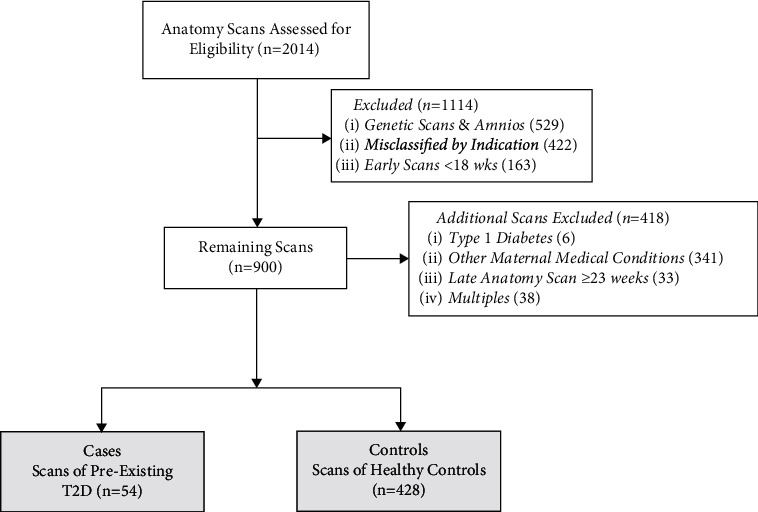
Methods: This was a case-control study (January 1, 2019 to December 31, 2019). Routine obstetrical ultrasounds performed between 18 and 22 weeks' gestation at a tertiary-care fetal assessment unit were reviewed. "Cases" comprised ultrasounds of fetuses exposed to pregestational T2D in utero. The control group was assembled from ultrasounds of healthy controls. Postprocessing measurements of fetal renal size and abdominal wall thickness from stored images were performed by two independent observers, and findings were compared between groups.
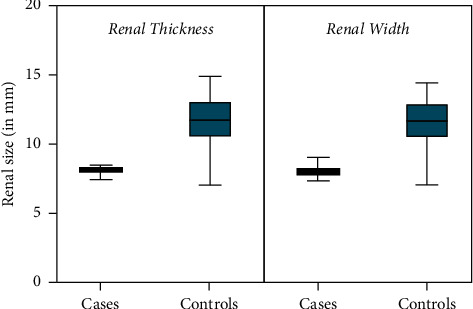
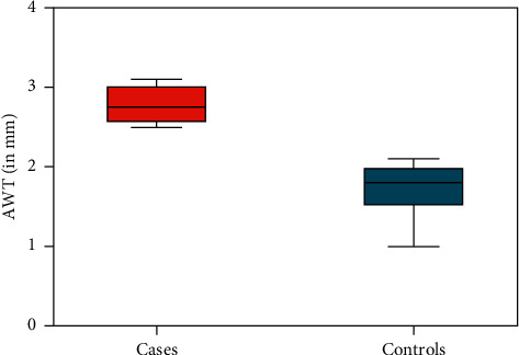
Results: There were 54 cases and 428 ultrasounds of healthy controls. The mean maternal age of cases was 32.1 years (SD 6.2) compared to 33.2 years (SD 5.3) for healthy controls, and the majority of ultrasounds were performed in multiparous patients (83%). At the 18 to 22 week ultrasound, there was a significant reduction in renal size amongst fetuses exposed to maternal T2D in utero compared to controls; among cases, the mean renal width was 8.0 mm (95% CI 7.8-8.1) compared to 11.4 mm (95% CI 10.6-12.7) in controls (p < 0.0001); the mean renal thickness among cases was 8.1 mm (95% CI 7.9-8.2) compared to 11.5 mm (95% CI 10.7-12.9) in controls (p=0.001). There was no obvious difference in estimated fetal weight between groups, yet fetuses exposed to maternal T2D had increased subcutaneous abdominal wall fat thickness at this early gestational age (p=0.008).
Conclusions: Fetal renal size in cases exposed to pregestational T2D is significantly smaller compared to controls, and subcutaneous abdominal wall fat is significantly thicker. Given emerging evidence about the developmental origins of disease, further study is needed to correlate the association between fetal renal size and fat distribution in the fetus and the long-term risk of chronic renal disease and diabetes in these offspring.
目的:探讨妊娠期2型糖尿病(T2D)暴露与常规产科超声检查胎儿肾脏大小和皮下脂肪厚度的关系。方法:病例对照研究(2019年1月1日至2019年12月31日)。常规产科超声在妊娠18和22周之间在一个三级保健胎儿评估单位进行审查。“病例”包括胎儿在子宫内暴露于妊娠期T2D的超声波。对照组由健康对照者的超声波组成。后处理测量胎儿肾脏大小和腹壁厚度从存储的图像进行了两个独立的观察员,并发现组之间的比较。结果:54例,正常对照428例。这些病例的平均母亲年龄为32.1岁(SD 6.2),而健康对照组为33.2岁(SD 5.3),大多数超声检查是在多胎患者中进行的(83%)。在18至22周的超声检查中,与对照组相比,子宫内暴露于母体T2D的胎儿肾脏大小显著减小;在这些病例中,平均肾脏宽度为8.0 mm (95% CI 7.8-8.1),而对照组为11.4 mm (95% CI 10.6-12.7) (p < 0.0001);病例的平均肾脏厚度为8.1 mm (95% CI 7.9-8.2),而对照组为11.5 mm (95% CI 10.7-12.9) (p=0.001)。两组之间胎儿体重的估计没有明显差异,但母体T2D暴露的胎儿在孕早期腹壁脂肪厚度增加(p=0.008)。结论:妊娠期暴露于T2D的胎儿肾脏大小明显小于对照组,皮下腹壁脂肪明显增厚。鉴于有关疾病发育起源的新证据,需要进一步研究胎儿肾脏大小和胎儿脂肪分布与这些后代患慢性肾脏疾病和糖尿病的长期风险之间的关系。
期刊介绍:
International Journal of Nephrology is a peer-reviewed, Open Access journal that publishes original research articles, review articles, and clinical studies focusing on the prevention, diagnosis, and management of kidney diseases and associated disorders. The journal welcomes submissions related to cell biology, developmental biology, genetics, immunology, pathology, pathophysiology of renal disease and progression, clinical nephrology, dialysis, and transplantation.

 求助内容:
求助内容: 应助结果提醒方式:


