Dingding Yang, Yan Zhao, Binbin Nie, Leiting An, Xiangdong Wan, Yazhou Wang, Wenting Wang, Guohong Cai, Shengxi Wu
{"title":"Progress in magnetic resonance imaging of autism model mice brain.","authors":"Dingding Yang, Yan Zhao, Binbin Nie, Leiting An, Xiangdong Wan, Yazhou Wang, Wenting Wang, Guohong Cai, Shengxi Wu","doi":"10.1002/wcs.1616","DOIUrl":null,"url":null,"abstract":"<p><p>Autism spectrum disorder (ASD) is a neurodevelopmental disease characterized by social disorder and stereotypical behaviors with an increasing incidence. ASD patients are suffering from varying degrees of mental retardation and language development abnormalities. Magnetic resonance imaging (MRI) is a noninvasive imaging technology to detect brain structural and functional dysfunction in vivo, playing an important role in the early diagnosisbasic research of ASD. High-field, small-animal MRI in basic research of autism model mice has provided a new approach to research the pathogenesis, characteristics, and intervention efficacy in autism. This article reviews MRI studies of mouse models of autism over the past 20 years. Reduced gray matter, abnormal connections of brain networks, and abnormal development of white matter fibers have been demonstrated in these studies, which are present in different proportions in the various mouse models. This provides a more macroscopic view for subsequent research on autism model mice. This article is categorized under: Cognitive Biology > Genes and Environment Neuroscience > Computation Neuroscience > Genes, Molecules, and Cells Neuroscience > Development.</p>","PeriodicalId":47720,"journal":{"name":"Wiley Interdisciplinary Reviews-Cognitive Science","volume":" ","pages":"e1616"},"PeriodicalIF":3.8000,"publicationDate":"2022-11-01","publicationTypes":"Journal Article","fieldsOfStudy":null,"isOpenAccess":false,"openAccessPdf":"","citationCount":"0","resultStr":null,"platform":"Semanticscholar","paperid":null,"PeriodicalName":"Wiley Interdisciplinary Reviews-Cognitive Science","FirstCategoryId":"102","ListUrlMain":"https://doi.org/10.1002/wcs.1616","RegionNum":2,"RegionCategory":"心理学","ArticlePicture":[],"TitleCN":null,"AbstractTextCN":null,"PMCID":null,"EPubDate":"2022/8/5 0:00:00","PubModel":"Epub","JCR":"Q1","JCRName":"PSYCHOLOGY, EXPERIMENTAL","Score":null,"Total":0}
引用次数: 0
Abstract
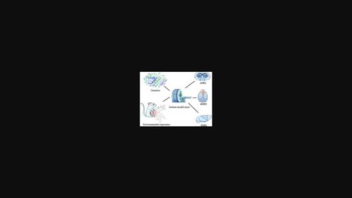
Autism spectrum disorder (ASD) is a neurodevelopmental disease characterized by social disorder and stereotypical behaviors with an increasing incidence. ASD patients are suffering from varying degrees of mental retardation and language development abnormalities. Magnetic resonance imaging (MRI) is a noninvasive imaging technology to detect brain structural and functional dysfunction in vivo, playing an important role in the early diagnosisbasic research of ASD. High-field, small-animal MRI in basic research of autism model mice has provided a new approach to research the pathogenesis, characteristics, and intervention efficacy in autism. This article reviews MRI studies of mouse models of autism over the past 20 years. Reduced gray matter, abnormal connections of brain networks, and abnormal development of white matter fibers have been demonstrated in these studies, which are present in different proportions in the various mouse models. This provides a more macroscopic view for subsequent research on autism model mice. This article is categorized under: Cognitive Biology > Genes and Environment Neuroscience > Computation Neuroscience > Genes, Molecules, and Cells Neuroscience > Development.

 求助内容:
求助内容: 应助结果提醒方式:


