{"title":"Myosin assembly of smooth muscle: from ribbons and side polarity to a row polar helical model.","authors":"Isabel J Sobieszek, Apolinary Sobieszek","doi":"10.1007/s10974-022-09622-4","DOIUrl":null,"url":null,"abstract":"<p><p>After decades of debate over the structure of smooth muscle myosin filaments, it is still unclear whether they are helical, as in all other muscle types, or square in shape. In both cases bipolar building units are proposed, but the deduced cross-bridge arrangements are fundamentally different. The opposite polarity of the adjusting longitudinal rows is proposed for the helical structure, while in the case of square filaments, or myosin ribbons, only their two faces are appositively polarized. Analysis of our unpublished archival data on light meromyosin (LMM) paracrystals and myosin rod assemblies as well as the filaments themselves indicated that the rods were assembled with a 6°-7° tilt angle from the rods' longitudinal axis, in contrast to the lack of tilt in LMM, both exhibiting a 14.3 nm myosin periodicity. Optical diffraction analysis of EM images of the rod assemblies and those of intact myosin confirmed their helical architecture characterized by 28 nm residue translations, 172 nm repeats and 516 nm pitch. A detailed helical model of these filaments was elucidated with bipolar tetramer building units made of two polar trimers. The filaments elongate at their two ends in a head-to-head manner, enabling targeted cross-bridge polarity of the adjacent rows, in the form of a unique Boerdijk-Coxeter type helix, similar to that of collagen or desmin fibers, with the covalent links replaced by a head-to-head clasp.</p>","PeriodicalId":16422,"journal":{"name":"Journal of Muscle Research and Cell Motility","volume":"43 3","pages":"113-133"},"PeriodicalIF":1.7000,"publicationDate":"2022-09-01","publicationTypes":"Journal Article","fieldsOfStudy":null,"isOpenAccess":false,"openAccessPdf":"https://www.ncbi.nlm.nih.gov/pmc/articles/PMC9420085/pdf/","citationCount":"3","resultStr":null,"platform":"Semanticscholar","paperid":null,"PeriodicalName":"Journal of Muscle Research and Cell Motility","FirstCategoryId":"99","ListUrlMain":"https://doi.org/10.1007/s10974-022-09622-4","RegionNum":3,"RegionCategory":"生物学","ArticlePicture":[],"TitleCN":null,"AbstractTextCN":null,"PMCID":null,"EPubDate":"2022/7/16 0:00:00","PubModel":"Epub","JCR":"Q4","JCRName":"CELL BIOLOGY","Score":null,"Total":0}
引用次数: 3
Abstract
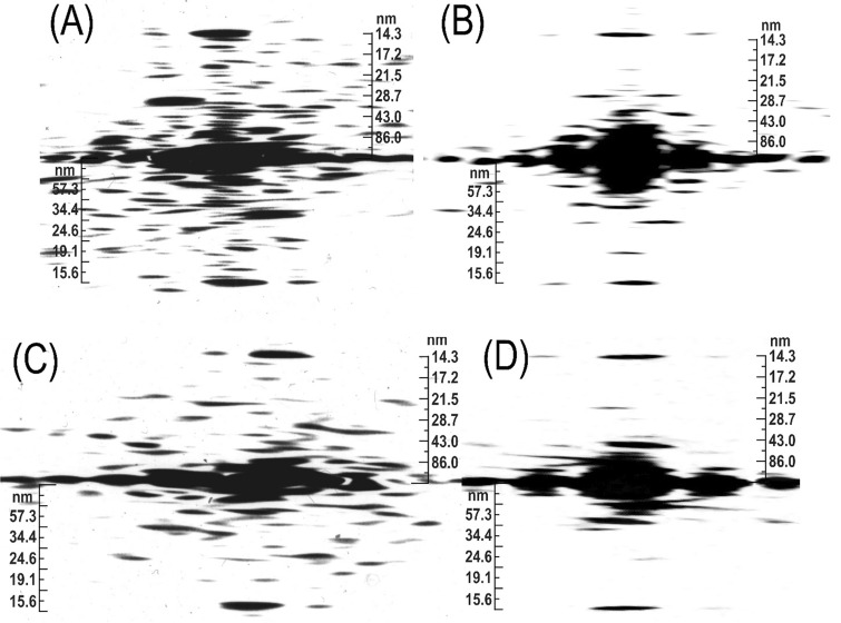
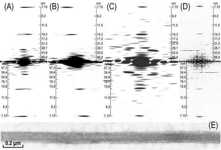
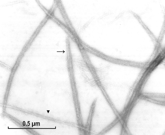
After decades of debate over the structure of smooth muscle myosin filaments, it is still unclear whether they are helical, as in all other muscle types, or square in shape. In both cases bipolar building units are proposed, but the deduced cross-bridge arrangements are fundamentally different. The opposite polarity of the adjusting longitudinal rows is proposed for the helical structure, while in the case of square filaments, or myosin ribbons, only their two faces are appositively polarized. Analysis of our unpublished archival data on light meromyosin (LMM) paracrystals and myosin rod assemblies as well as the filaments themselves indicated that the rods were assembled with a 6°-7° tilt angle from the rods' longitudinal axis, in contrast to the lack of tilt in LMM, both exhibiting a 14.3 nm myosin periodicity. Optical diffraction analysis of EM images of the rod assemblies and those of intact myosin confirmed their helical architecture characterized by 28 nm residue translations, 172 nm repeats and 516 nm pitch. A detailed helical model of these filaments was elucidated with bipolar tetramer building units made of two polar trimers. The filaments elongate at their two ends in a head-to-head manner, enabling targeted cross-bridge polarity of the adjacent rows, in the form of a unique Boerdijk-Coxeter type helix, similar to that of collagen or desmin fibers, with the covalent links replaced by a head-to-head clasp.
期刊介绍:
The Journal of Muscle Research and Cell Motility has as its main aim the publication of original research which bears on either the excitation and contraction of muscle, the analysis of any one of the processes involved therein, the processes underlying contractility and motility of animal and plant cells, the toxicology and pharmacology related to contractility, or the formation, dynamics and turnover of contractile structures in muscle and non-muscle cells. Studies describing the impact of pathogenic mutations in genes encoding components of contractile structures in humans or animals are welcome, provided they offer mechanistic insight into the disease process or the underlying gene function. The policy of the Journal is to encourage any form of novel practical study whatever its specialist interest, as long as it falls within this broad field. Theoretical essays are welcome provided that they are concise and suggest practical ways in which they may be tested. Manuscripts reporting new mutations in known disease genes without validation and mechanistic insight will not be considered. It is the policy of the journal that cells lines, hybridomas and DNA clones should be made available by the developers to any qualified investigator. Submission of a manuscript for publication constitutes an agreement of the authors to abide by this principle.

 求助内容:
求助内容: 应助结果提醒方式:


