{"title":"Light and electron microscopic morphology of the fertilized egg and fertilized egg envelope of Poropanchax normani, Poeciliidae, Teleostei","authors":"Dong Heui Kim","doi":"10.1186/s42649-022-00075-0","DOIUrl":null,"url":null,"abstract":"<div><p>We examined the morphology of the fertilized egg and the fine structure of fertilized egg envelopes of <i>Poropanchax normani</i> belonging to the family Poeciliidae, also known as Norman’s lampeye using light and electron microscopes. The fertilized eggs with narrow perivitelline space were found to be spherical and demersal, additionally containing small oil droplets in the vitelline membrane. Further, a bundle of adhesive filaments was observed to be present on one side of the fertilized egg. These filaments possessed remarkably high elasticity and were approximately 1-3 mm in length. The size of the fertilized egg was determined to be about 1.49 ± 0.07 mm (<i>n</i> = 30). The outer surface appeared smooth, and adhesive filaments originating at different location of the surface of the envelope were found to be distributed around the egg envelope and were joined together to form a single long bundle in scanning electron microscopic observation. A peak-like structure formed of several straight wrinkles was observed around the micropyle. However, the complete structure of the micropyle could not be studied due to the depth at which it was located. Additionally, the total thickness of the egg envelope was ascertained to be approximately12.5–14.5 μm. The egg envelope consisted of two distinct layers, an outer electron dense layer and an inner lamellar layer, further consisting of 10 sublayers of varying thicknesses. Collectively, it was observed that the morphological characteristics of the fertilized egg, fine structures surrounding the micropyle, outer surface, adhesive structure consisting adhesive filaments, and sections of fertilized egg envelope displayed species specificity.</p></div>","PeriodicalId":470,"journal":{"name":"Applied Microscopy","volume":"52 1","pages":""},"PeriodicalIF":0.0000,"publicationDate":"2022-07-13","publicationTypes":"Journal Article","fieldsOfStudy":null,"isOpenAccess":false,"openAccessPdf":"https://www.ncbi.nlm.nih.gov/pmc/articles/PMC9279538/pdf/","citationCount":"0","resultStr":null,"platform":"Semanticscholar","paperid":null,"PeriodicalName":"Applied Microscopy","FirstCategoryId":"1085","ListUrlMain":"https://link.springer.com/article/10.1186/s42649-022-00075-0","RegionNum":0,"RegionCategory":null,"ArticlePicture":[],"TitleCN":null,"AbstractTextCN":null,"PMCID":null,"EPubDate":"","PubModel":"","JCR":"Q3","JCRName":"Immunology and Microbiology","Score":null,"Total":0}
引用次数: 0
Abstract
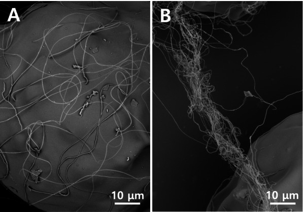
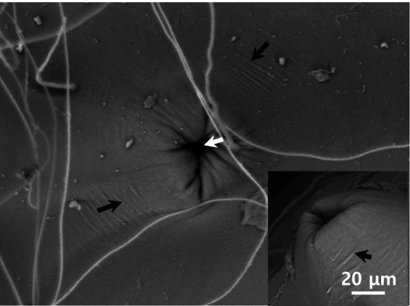
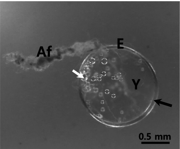
We examined the morphology of the fertilized egg and the fine structure of fertilized egg envelopes of Poropanchax normani belonging to the family Poeciliidae, also known as Norman’s lampeye using light and electron microscopes. The fertilized eggs with narrow perivitelline space were found to be spherical and demersal, additionally containing small oil droplets in the vitelline membrane. Further, a bundle of adhesive filaments was observed to be present on one side of the fertilized egg. These filaments possessed remarkably high elasticity and were approximately 1-3 mm in length. The size of the fertilized egg was determined to be about 1.49 ± 0.07 mm (n = 30). The outer surface appeared smooth, and adhesive filaments originating at different location of the surface of the envelope were found to be distributed around the egg envelope and were joined together to form a single long bundle in scanning electron microscopic observation. A peak-like structure formed of several straight wrinkles was observed around the micropyle. However, the complete structure of the micropyle could not be studied due to the depth at which it was located. Additionally, the total thickness of the egg envelope was ascertained to be approximately12.5–14.5 μm. The egg envelope consisted of two distinct layers, an outer electron dense layer and an inner lamellar layer, further consisting of 10 sublayers of varying thicknesses. Collectively, it was observed that the morphological characteristics of the fertilized egg, fine structures surrounding the micropyle, outer surface, adhesive structure consisting adhesive filaments, and sections of fertilized egg envelope displayed species specificity.
Applied MicroscopyImmunology and Microbiology-Applied Microbiology and Biotechnology
CiteScore
3.40
自引率
0.00%
发文量
10
审稿时长
10 weeks
期刊介绍:
Applied Microscopy is a peer-reviewed journal sponsored by the Korean Society of Microscopy. The journal covers all the interdisciplinary fields of technological developments in new microscopy methods and instrumentation and their applications to biological or materials science for determining structure and chemistry. ISSN: 22875123, 22874445.
 求助内容:
求助内容: 应助结果提醒方式:


