Marek Duczkowski, Agnieszka Duczkowska, Elżbieta Michalak, Agnieszka Tomasik-Kowalska, Sylwia Szkudlińska-Pawlak, Bartosz Pachuta, Anna Raciborska, Monika Bekiesińska-Figatowska
{"title":"Suspicion of Recurrent Osteosarcoma in a Patient with Pseudotumour Adjacent To the Endoprosthesis.","authors":"Marek Duczkowski, Agnieszka Duczkowska, Elżbieta Michalak, Agnieszka Tomasik-Kowalska, Sylwia Szkudlińska-Pawlak, Bartosz Pachuta, Anna Raciborska, Monika Bekiesińska-Figatowska","doi":"10.15557/JoU.2022.0023","DOIUrl":null,"url":null,"abstract":"<p><strong>Purpose: </strong>Pseudotumor is a rare complication after arthroplasty, most often of the hip joint, in response to metal particles present in the implant. There are merely sporadic reports of pseudotumor in patients with bone sarcoma after sparing surgery with endoprosthesis implant. The aim of this study is to present the characteristic imaging features of pseudotumor.</p><p><strong>Case report: </strong>We present a case of a 21-year-old male patient in whom a scheduled follow-up ultrasound revealed a painless lesion suspected of local recurrence at the border of the endoprosthesis and the bone stump 3.5 years after the end of treatment for osteosarcoma of the femur. Histopathology of the biopsy specimen revealed that the lesion was a pseudotumor.</p><p><strong>Conclusions: </strong>Although pseudotumor is sporadic in patients treated with endoprosthesis for bone sarcoma, their prolonged survival could bear the risk of such a complication. Imaging studies, in particular ultrasound, may be helpful in differentiating from local recurrence of sarcoma, however, the histopathology of the specimen obtained by open biopsy at a reference center is crucial for the final diagnosis.</p>","PeriodicalId":45612,"journal":{"name":"Journal of Ultrasonography","volume":"22 89","pages":"140-143"},"PeriodicalIF":1.3000,"publicationDate":"2022-04-27","publicationTypes":"Journal Article","fieldsOfStudy":null,"isOpenAccess":false,"openAccessPdf":"https://ftp.ncbi.nlm.nih.gov/pub/pmc/oa_pdf/7d/2d/jou-22-140.PMC9231517.pdf","citationCount":"0","resultStr":null,"platform":"Semanticscholar","paperid":null,"PeriodicalName":"Journal of Ultrasonography","FirstCategoryId":"1085","ListUrlMain":"https://doi.org/10.15557/JoU.2022.0023","RegionNum":0,"RegionCategory":null,"ArticlePicture":[],"TitleCN":null,"AbstractTextCN":null,"PMCID":null,"EPubDate":"2022/4/1 0:00:00","PubModel":"eCollection","JCR":"Q3","JCRName":"RADIOLOGY, NUCLEAR MEDICINE & MEDICAL IMAGING","Score":null,"Total":0}
引用次数: 0
Abstract
Purpose: Pseudotumor is a rare complication after arthroplasty, most often of the hip joint, in response to metal particles present in the implant. There are merely sporadic reports of pseudotumor in patients with bone sarcoma after sparing surgery with endoprosthesis implant. The aim of this study is to present the characteristic imaging features of pseudotumor.
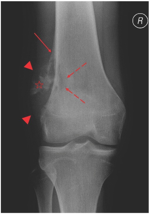
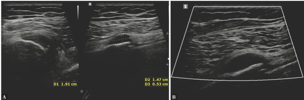
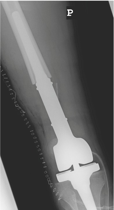
Case report: We present a case of a 21-year-old male patient in whom a scheduled follow-up ultrasound revealed a painless lesion suspected of local recurrence at the border of the endoprosthesis and the bone stump 3.5 years after the end of treatment for osteosarcoma of the femur. Histopathology of the biopsy specimen revealed that the lesion was a pseudotumor.
Conclusions: Although pseudotumor is sporadic in patients treated with endoprosthesis for bone sarcoma, their prolonged survival could bear the risk of such a complication. Imaging studies, in particular ultrasound, may be helpful in differentiating from local recurrence of sarcoma, however, the histopathology of the specimen obtained by open biopsy at a reference center is crucial for the final diagnosis.

 求助内容:
求助内容: 应助结果提醒方式:


