{"title":"Characterizations of alveolar repair after mandibular second molar extraction: an experimental study in rats.","authors":"Jianbin Li, Zhenxian Sheng, Jing Sun, Ronglin Wang, Xijiao Yu","doi":"10.1590/1678-7757-2022-0010","DOIUrl":null,"url":null,"abstract":"<p><strong>Background: </strong>Characterizations of rat mandibular second molar extraction socket with significantly different buccal and lingual alveolar ridge width remain unclear.</p><p><strong>Objective: </strong>To observe alterations in the alveolar ridge after extraction of mandibular second molars, and to examine processes of alveolar socket healing in an experimental model of alveolar ridge absorption and preservation.</p><p><strong>Methodology: </strong>Eighteen Wistar rats were included and divided into six groups regarding healing time in the study. Bilateral mandibular second molars were extracted. The rats with tooth extraction sockets took 0, 1.5, 2, 3, 4 and 8 weeks of healing. Histological observation, tartrate-resistant acidic phosphatase (TRAP) staining, Masson's trichrome staining, immunohistochemical staining and micro-computed tomography (micro-CT) were applied to estimate alterations in the alveolar ridge.</p><p><strong>Results: </strong>Different buccal and lingual alveolar ridge width led to different height loss. Lingual wall height (LH) decreased significantly two weeks after tooth extraction. Buccal wall height rarely reduced its higher ridge width. From two to eight weeks after extraction, bone volume (BV/TV), density (BMD), and trabecular thickness (Tb.Th) progressively increased in the alveolar socket, which gradually decreased in Tb.Sp and Tb.N. LH showed no significant change during the same period. Osteogenic marker OCN and OPN increased during bone repair from two to eight weeks. The reduced height of the lingual wall of the tooth extraction socket was rarely repaired in the later repair stage. Osteoclast activity led to absorption of the alveolar ridge of the alveolar bone wall within two weeks after operation. We observed positive expression of EMMPRIN and MMP-9 in osteoclasts that participated in the absorption of the spire region.</p><p><strong>Conclusion: </strong>Extraction of rat mandibular second molars may help the study of alveolar ridge absorption and preservation. The EMMPRIN-MMP-9 pathway may be a candidate for further study on attenuating bone resorption after tooth extraction.</p>","PeriodicalId":321675,"journal":{"name":"Journal of applied oral science : revista FOB","volume":" ","pages":"e20220010"},"PeriodicalIF":0.0000,"publicationDate":"2022-07-08","publicationTypes":"Journal Article","fieldsOfStudy":null,"isOpenAccess":false,"openAccessPdf":"https://www.ncbi.nlm.nih.gov/pmc/articles/PMC9275398/pdf/","citationCount":"3","resultStr":null,"platform":"Semanticscholar","paperid":null,"PeriodicalName":"Journal of applied oral science : revista FOB","FirstCategoryId":"3","ListUrlMain":"https://doi.org/10.1590/1678-7757-2022-0010","RegionNum":0,"RegionCategory":null,"ArticlePicture":[],"TitleCN":null,"AbstractTextCN":null,"PMCID":null,"EPubDate":"2022/1/1 0:00:00","PubModel":"eCollection","JCR":"","JCRName":"","Score":null,"Total":0}
引用次数: 3
Abstract
Background: Characterizations of rat mandibular second molar extraction socket with significantly different buccal and lingual alveolar ridge width remain unclear.
Objective: To observe alterations in the alveolar ridge after extraction of mandibular second molars, and to examine processes of alveolar socket healing in an experimental model of alveolar ridge absorption and preservation.
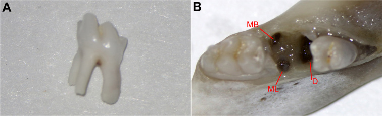
Methodology: Eighteen Wistar rats were included and divided into six groups regarding healing time in the study. Bilateral mandibular second molars were extracted. The rats with tooth extraction sockets took 0, 1.5, 2, 3, 4 and 8 weeks of healing. Histological observation, tartrate-resistant acidic phosphatase (TRAP) staining, Masson's trichrome staining, immunohistochemical staining and micro-computed tomography (micro-CT) were applied to estimate alterations in the alveolar ridge.
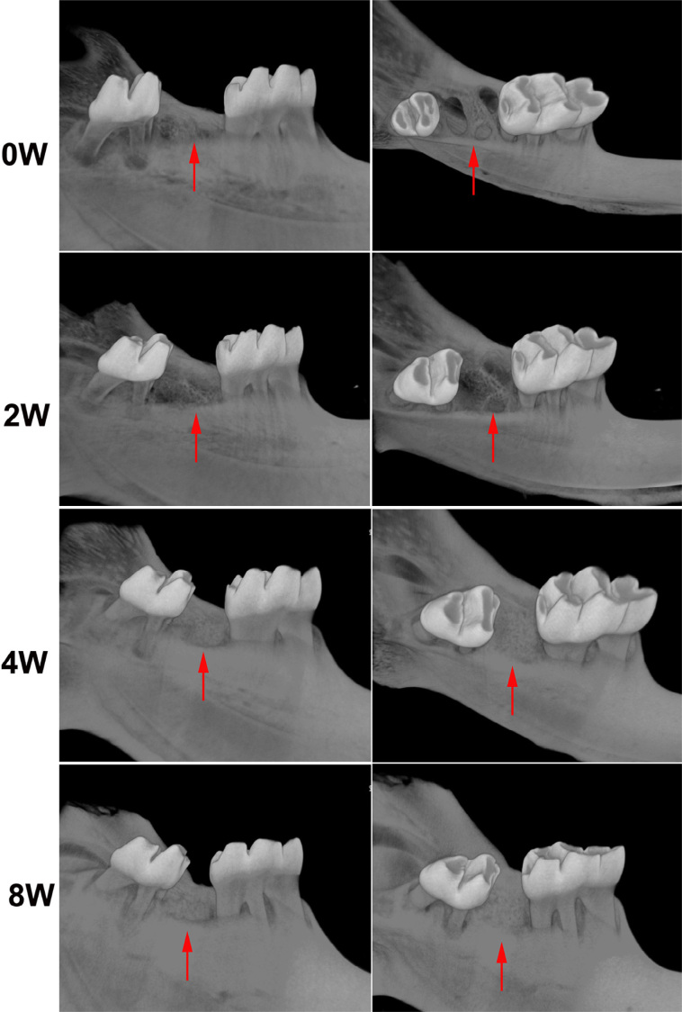
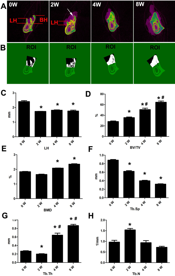
Results: Different buccal and lingual alveolar ridge width led to different height loss. Lingual wall height (LH) decreased significantly two weeks after tooth extraction. Buccal wall height rarely reduced its higher ridge width. From two to eight weeks after extraction, bone volume (BV/TV), density (BMD), and trabecular thickness (Tb.Th) progressively increased in the alveolar socket, which gradually decreased in Tb.Sp and Tb.N. LH showed no significant change during the same period. Osteogenic marker OCN and OPN increased during bone repair from two to eight weeks. The reduced height of the lingual wall of the tooth extraction socket was rarely repaired in the later repair stage. Osteoclast activity led to absorption of the alveolar ridge of the alveolar bone wall within two weeks after operation. We observed positive expression of EMMPRIN and MMP-9 in osteoclasts that participated in the absorption of the spire region.
Conclusion: Extraction of rat mandibular second molars may help the study of alveolar ridge absorption and preservation. The EMMPRIN-MMP-9 pathway may be a candidate for further study on attenuating bone resorption after tooth extraction.

 求助内容:
求助内容: 应助结果提醒方式:


