Mohit Jambhulkar, Jasvinder K Bhatia, Samresh K Singh
{"title":"Correlation Between Fine-Needle Aspiration Cytology, Cell Block Cytology, and Histopathology in the Diagnosis of Thyroid Lesions.","authors":"Mohit Jambhulkar, Jasvinder K Bhatia, Samresh K Singh","doi":"10.4103/joc.joc_80_21","DOIUrl":null,"url":null,"abstract":"<p><strong>Context: </strong>Fine-needle aspiration cytology is considered the gold standard screening test in the evaluation of a thyroid nodule. We studied whether cell block cytology can be used in addition to conventional smears for the evaluation of tissue from fine-needle aspirations or fluid aspirations and also compared it with histopathological diagnosis.</p><p><strong>Aims: </strong>The primary aim of this study was to know the utility of cell blocks in the diagnosis of thyroid lesions.</p><p><strong>Settings and design: </strong>This was a prospective observational study conducted from June 2018 to September 2020 at a tertiary Care Hospital in Eastern India. Ethical approval was obtained from the Ethics Committee of the institution. Patients above 18 years who presented with goiter were included in the study. Thirty patients were enrolled in the study after informed consent.</p><p><strong>Methods and material: </strong>Smears prepared from the aspirates were stained with Leishman-Giemsa (LG) and Pap stain. The remnant from the needle hub was transferred to a sterile container. Cell blocks were prepared from the remnants. Smears were scored based on cell obscuration by blood, cellularity, cell degeneration, and cell architecture. The results were compared with histopathology.</p><p><strong>Statistical analysis used: </strong>Data were recorded using Microsoft Excel. Descriptive statistics, frequency, and proportion were used to describe demographic variables.</p><p><strong>Results: </strong>The majority of the patients (23.3%) were in their third decade of life, followed by 16.7% of the patients in their fourth and fifth decades. The patient age ranged from 25 to 80 years, with a mean age of 50.83 years and a standard deviation of 16.72. The largest number of patients were females accounting for 80% (24/30) of the total participants. The majority of the patients (36.7%) (11/30) had thyroid gland enlargement for a period of 15 days to three months. 14% of the participants were not able to recall its duration. The majority (60%) (18/30) had left lobe lesions, followed by 33.3% (10/30) who had right lobe lesions, and 6.7% (2/30) who had bilateral lobe swelling. The mean size of the lesion was 2.84 cm. 50% were found to be Bethesda II lesions, while 13.3% were Bethesda IV, and 36.7% were found to be Bethesda VI lesions. The cell block score (7) was found to be better compared to Fine Needle Aspiration Cytology (FNAC) (4.7). Tissue Coagulum Clot and Clot Scrape methods were found to yield better results compared to the Cytocentrifuge method. The <i>P</i> value was found to be significant (<0.001).</p><p><strong>Conclusions: </strong>Cell blocks were found to improve the cell morphology compared to FNAC alone and can be used as an adjunct to FNAC in the diagnosis of various thyroid lesions.</p>","PeriodicalId":50217,"journal":{"name":"Journal of Cytology","volume":"39 3","pages":"91-97"},"PeriodicalIF":1.0000,"publicationDate":"2022-07-01","publicationTypes":"Journal Article","fieldsOfStudy":null,"isOpenAccess":false,"openAccessPdf":"https://www.ncbi.nlm.nih.gov/pmc/articles/PMC9585813/pdf/","citationCount":"1","resultStr":null,"platform":"Semanticscholar","paperid":null,"PeriodicalName":"Journal of Cytology","FirstCategoryId":"3","ListUrlMain":"https://doi.org/10.4103/joc.joc_80_21","RegionNum":4,"RegionCategory":"医学","ArticlePicture":[],"TitleCN":null,"AbstractTextCN":null,"PMCID":null,"EPubDate":"2022/7/30 0:00:00","PubModel":"Epub","JCR":"Q4","JCRName":"MEDICAL LABORATORY TECHNOLOGY","Score":null,"Total":0}
引用次数: 1
Abstract
Context: Fine-needle aspiration cytology is considered the gold standard screening test in the evaluation of a thyroid nodule. We studied whether cell block cytology can be used in addition to conventional smears for the evaluation of tissue from fine-needle aspirations or fluid aspirations and also compared it with histopathological diagnosis.
Aims: The primary aim of this study was to know the utility of cell blocks in the diagnosis of thyroid lesions.
Settings and design: This was a prospective observational study conducted from June 2018 to September 2020 at a tertiary Care Hospital in Eastern India. Ethical approval was obtained from the Ethics Committee of the institution. Patients above 18 years who presented with goiter were included in the study. Thirty patients were enrolled in the study after informed consent.
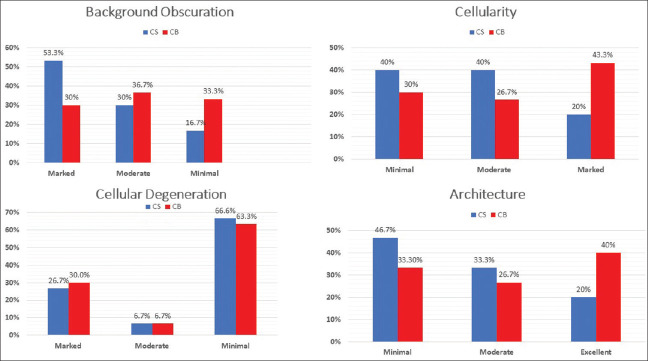
Methods and material: Smears prepared from the aspirates were stained with Leishman-Giemsa (LG) and Pap stain. The remnant from the needle hub was transferred to a sterile container. Cell blocks were prepared from the remnants. Smears were scored based on cell obscuration by blood, cellularity, cell degeneration, and cell architecture. The results were compared with histopathology.
Statistical analysis used: Data were recorded using Microsoft Excel. Descriptive statistics, frequency, and proportion were used to describe demographic variables.
Results: The majority of the patients (23.3%) were in their third decade of life, followed by 16.7% of the patients in their fourth and fifth decades. The patient age ranged from 25 to 80 years, with a mean age of 50.83 years and a standard deviation of 16.72. The largest number of patients were females accounting for 80% (24/30) of the total participants. The majority of the patients (36.7%) (11/30) had thyroid gland enlargement for a period of 15 days to three months. 14% of the participants were not able to recall its duration. The majority (60%) (18/30) had left lobe lesions, followed by 33.3% (10/30) who had right lobe lesions, and 6.7% (2/30) who had bilateral lobe swelling. The mean size of the lesion was 2.84 cm. 50% were found to be Bethesda II lesions, while 13.3% were Bethesda IV, and 36.7% were found to be Bethesda VI lesions. The cell block score (7) was found to be better compared to Fine Needle Aspiration Cytology (FNAC) (4.7). Tissue Coagulum Clot and Clot Scrape methods were found to yield better results compared to the Cytocentrifuge method. The P value was found to be significant (<0.001).
Conclusions: Cell blocks were found to improve the cell morphology compared to FNAC alone and can be used as an adjunct to FNAC in the diagnosis of various thyroid lesions.
期刊介绍:
The Journal of Cytology is the official Quarterly publication of the Indian Academy of Cytologists. It is in the 25th year of publication in the year 2008. The journal covers all aspects of diagnostic cytology, including fine needle aspiration cytology, gynecological and non-gynecological cytology. Articles on ancillary techniques, like cytochemistry, immunocytochemistry, electron microscopy, molecular cytopathology, as applied to cytological material are also welcome. The journal gives preference to clinically oriented studies over experimental and animal studies. The Journal would publish peer-reviewed original research papers, case reports, systematic reviews, meta-analysis, and debates.
 求助内容:
求助内容: 应助结果提醒方式:


