{"title":"Brain Region and Sex-specific Changes in Mitochondrial Biogenesis Induced by Acute Trimethyltin Exposure.","authors":"Jung Ho Lee, Eun Hye Jang, Soon Ae Kim","doi":"10.9758/cpn.2022.20.3.474","DOIUrl":null,"url":null,"abstract":"<p><strong>Objective: </strong>In this study, we investigated sex- and region-specific effects of acute trimethyltin (TMT) exposure on mitochondrial biogenesis.</p><p><strong>Methods: </strong>We treated TMT to primary neuronal cultures and 4-week-old male and female mice. We measured the mitochondrial DNA copy numbers using the quantitative polymerase chain reaction method. We also measured mitochondrial biogenesis related genes (sirtuin-1, estrogen-related receptor alpha, cytochrome C oxidase subunit IV) by western blotting.</p><p><strong>Results: </strong>The mitochondrial DNA copy number increased in the primary hippocampal neuron; however, it decreased in the primary cortical neuron. The mitochondrial copy number increased in the hippocampus and decreased in the cortex in the TMT treated female mice, though the mitochondrial copy number increased in both cortex and hippocampus in the TMT treated male mice. TMT treatment increased sirtuin-1 expression in the male hippocampus but did not in the female brain. In the female brain, estrogen-related receptor alpha expression decreased in the cortex though there is no significant change in the male brain. The protein level of mitochondrial protein, cytochrome C oxidase subunit IV, increased in both cortex and hippocampus after TMT injection in male mice brain, but not in female mice brain.</p><p><strong>Conclusion: </strong>Our data suggest that acute TMT exposure induces distinct sex-specific metabolic characteristics in the brain before significant sexual maturation.</p>","PeriodicalId":10420,"journal":{"name":"Clinical Psychopharmacology and Neuroscience","volume":"20 3","pages":"474-481"},"PeriodicalIF":2.4000,"publicationDate":"2022-08-31","publicationTypes":"Journal Article","fieldsOfStudy":null,"isOpenAccess":false,"openAccessPdf":"https://ftp.ncbi.nlm.nih.gov/pub/pmc/oa_pdf/bb/dd/cpn-20-3-474.PMC9329116.pdf","citationCount":"0","resultStr":null,"platform":"Semanticscholar","paperid":null,"PeriodicalName":"Clinical Psychopharmacology and Neuroscience","FirstCategoryId":"3","ListUrlMain":"https://doi.org/10.9758/cpn.2022.20.3.474","RegionNum":4,"RegionCategory":"医学","ArticlePicture":[],"TitleCN":null,"AbstractTextCN":null,"PMCID":null,"EPubDate":"","PubModel":"","JCR":"Q3","JCRName":"NEUROSCIENCES","Score":null,"Total":0}
引用次数: 0
Abstract
Objective: In this study, we investigated sex- and region-specific effects of acute trimethyltin (TMT) exposure on mitochondrial biogenesis.
Methods: We treated TMT to primary neuronal cultures and 4-week-old male and female mice. We measured the mitochondrial DNA copy numbers using the quantitative polymerase chain reaction method. We also measured mitochondrial biogenesis related genes (sirtuin-1, estrogen-related receptor alpha, cytochrome C oxidase subunit IV) by western blotting.
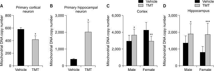
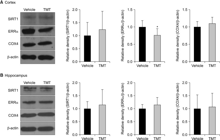
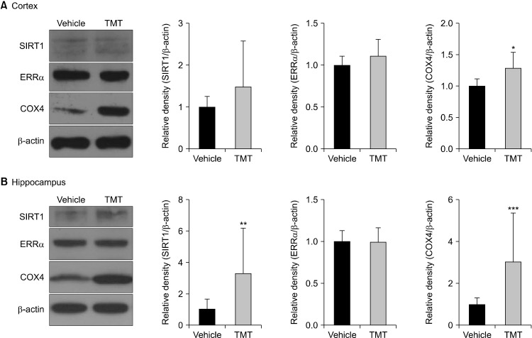
Results: The mitochondrial DNA copy number increased in the primary hippocampal neuron; however, it decreased in the primary cortical neuron. The mitochondrial copy number increased in the hippocampus and decreased in the cortex in the TMT treated female mice, though the mitochondrial copy number increased in both cortex and hippocampus in the TMT treated male mice. TMT treatment increased sirtuin-1 expression in the male hippocampus but did not in the female brain. In the female brain, estrogen-related receptor alpha expression decreased in the cortex though there is no significant change in the male brain. The protein level of mitochondrial protein, cytochrome C oxidase subunit IV, increased in both cortex and hippocampus after TMT injection in male mice brain, but not in female mice brain.
Conclusion: Our data suggest that acute TMT exposure induces distinct sex-specific metabolic characteristics in the brain before significant sexual maturation.
期刊介绍:
Clinical Psychopharmacology and Neuroscience (Clin Psychopharmacol Neurosci) launched in 2003, is the official journal of The Korean College of Neuropsychopharmacology (KCNP), and the associate journal for Asian College of Neuropsychopharmacology (AsCNP). This journal aims to publish evidence-based, scientifically written articles related to clinical and preclinical studies in the field of psychopharmacology and neuroscience. This journal intends to foster and encourage communications between psychiatrist, neuroscientist and all related experts in Asia as well as worldwide. It is published four times a year at the last day of February, May, August, and November.
 求助内容:
求助内容: 应助结果提醒方式:


