Magnifying Chromoendoscopy with Flexible Spectral Imaging Color Enhancement, Indigo Carmine, and Crystal Violet in Predicting the Histopathology of Colorectal Polyps: Diagnostic Value in a Scare-Setting Resource.
Nguyen Binh Pham, Khanh Truong Vu, Nam Hoai Nguyen, Ha Thi-Ngoc Doan, Thanh Trung Tran
{"title":"Magnifying Chromoendoscopy with Flexible Spectral Imaging Color Enhancement, Indigo Carmine, and Crystal Violet in Predicting the Histopathology of Colorectal Polyps: Diagnostic Value in a Scare-Setting Resource.","authors":"Nguyen Binh Pham, Khanh Truong Vu, Nam Hoai Nguyen, Ha Thi-Ngoc Doan, Thanh Trung Tran","doi":"10.1155/2022/6402904","DOIUrl":null,"url":null,"abstract":"<p><strong>Background and aims: </strong>Virtual magnifying chromoendoscopy with flexible spectral imaging color enhancement (FICE), image-enhanced endoscopy techniques, and dye-staining magnifying chromoendoscopy (with Indigo carmine and Crystal violet) have contributed to better visualization of the pit pattern and vascular structure of colorectal polyp. Therefore, magnifying chromoendoscopy is capable of predicting the histopathological results of colorectal polyp without biopsy and remains their diagnostic values over time, especially in scare-setting resources. This study compared the images of magnifying chromoendoscopy between FICE, Indigo carmine, and Crystal violet and then assessed their diagnostic values based on colorectal polyps' histopathology as a gold standard.</p><p><strong>Methods: </strong>A total of 332 polyps of 266 patients were endoscopically evaluated from June 2016 to September 2019. After identified by white light endoscopy, polyps continued to be evaluated by virtual magnifying chromoendoscopy (×50-150 times) with FICE. The capillary-vessel pattern was divided into 5 subtypes according to the number, morphology, and distribution of the fine blood vessels according to Teixeira classification. Next, they were stained with Indigo carmine 0.2% and then Crystal violet 0.05% and were classified according to Kudo's pit pattern classification. Finally, polyps were resected by endoscopy or surgery and biopsy and compared with histopathological results of either neoplastic or nonplastic polyp.</p><p><strong>Results: </strong>The number of neoplastic polyps was 278/332 with 231 adenoma polyps and 47 carcinoma polyps. Magnifying chromoendoscopy has high sensitivity and accuracy when compared with the histopathological results of colorectal polyps. The sensitivity, specificity, and accuracy of magnifying chromoendoscopy with Crystal violet are 97.2%, 72.2%, and 93.0%; with Indigo carmine are 96.0%, 72.2%, and 92.1%; and with FICE are 92.1%, 68.5%, and 88.3%.</p><p><strong>Conclusions: </strong>Among the three methods, Crystal violet has the highest sensitivity and accuracy in predicting histopathological results of colorectal polyps. FICE has shown its diagnostic value with reliable sensitivity and accuracy and should still be a reasonable endoscopic choice for physicians in scare-setting resources regardless its moderate specificity. Physicians should base on their facility and capability to determine an appropriate endoscopy technique.</p>","PeriodicalId":12597,"journal":{"name":"Gastroenterology Research and Practice","volume":" ","pages":"6402904"},"PeriodicalIF":2.0000,"publicationDate":"2022-07-13","publicationTypes":"Journal Article","fieldsOfStudy":null,"isOpenAccess":false,"openAccessPdf":"https://www.ncbi.nlm.nih.gov/pmc/articles/PMC9300359/pdf/","citationCount":"1","resultStr":null,"platform":"Semanticscholar","paperid":null,"PeriodicalName":"Gastroenterology Research and Practice","FirstCategoryId":"3","ListUrlMain":"https://doi.org/10.1155/2022/6402904","RegionNum":4,"RegionCategory":"医学","ArticlePicture":[],"TitleCN":null,"AbstractTextCN":null,"PMCID":null,"EPubDate":"2022/1/1 0:00:00","PubModel":"eCollection","JCR":"Q3","JCRName":"GASTROENTEROLOGY & HEPATOLOGY","Score":null,"Total":0}
引用次数: 1
Abstract
Background and aims: Virtual magnifying chromoendoscopy with flexible spectral imaging color enhancement (FICE), image-enhanced endoscopy techniques, and dye-staining magnifying chromoendoscopy (with Indigo carmine and Crystal violet) have contributed to better visualization of the pit pattern and vascular structure of colorectal polyp. Therefore, magnifying chromoendoscopy is capable of predicting the histopathological results of colorectal polyp without biopsy and remains their diagnostic values over time, especially in scare-setting resources. This study compared the images of magnifying chromoendoscopy between FICE, Indigo carmine, and Crystal violet and then assessed their diagnostic values based on colorectal polyps' histopathology as a gold standard.
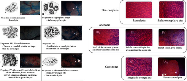
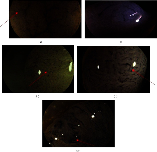
Methods: A total of 332 polyps of 266 patients were endoscopically evaluated from June 2016 to September 2019. After identified by white light endoscopy, polyps continued to be evaluated by virtual magnifying chromoendoscopy (×50-150 times) with FICE. The capillary-vessel pattern was divided into 5 subtypes according to the number, morphology, and distribution of the fine blood vessels according to Teixeira classification. Next, they were stained with Indigo carmine 0.2% and then Crystal violet 0.05% and were classified according to Kudo's pit pattern classification. Finally, polyps were resected by endoscopy or surgery and biopsy and compared with histopathological results of either neoplastic or nonplastic polyp.
Results: The number of neoplastic polyps was 278/332 with 231 adenoma polyps and 47 carcinoma polyps. Magnifying chromoendoscopy has high sensitivity and accuracy when compared with the histopathological results of colorectal polyps. The sensitivity, specificity, and accuracy of magnifying chromoendoscopy with Crystal violet are 97.2%, 72.2%, and 93.0%; with Indigo carmine are 96.0%, 72.2%, and 92.1%; and with FICE are 92.1%, 68.5%, and 88.3%.
Conclusions: Among the three methods, Crystal violet has the highest sensitivity and accuracy in predicting histopathological results of colorectal polyps. FICE has shown its diagnostic value with reliable sensitivity and accuracy and should still be a reasonable endoscopic choice for physicians in scare-setting resources regardless its moderate specificity. Physicians should base on their facility and capability to determine an appropriate endoscopy technique.
期刊介绍:
Gastroenterology Research and Practice is a peer-reviewed, Open Access journal which publishes original research articles, review articles and clinical studies based on all areas of gastroenterology, hepatology, pancreas and biliary, and related cancers. The journal welcomes submissions on the physiology, pathophysiology, etiology, diagnosis and therapy of gastrointestinal diseases. The aim of the journal is to provide cutting edge research related to the field of gastroenterology, as well as digestive diseases and disorders.
Topics of interest include:
Management of pancreatic diseases
Third space endoscopy
Endoscopic resection
Therapeutic endoscopy
Therapeutic endosonography.
 求助内容:
求助内容: 应助结果提醒方式:


