Anna Lardone, Marianna Liparoti, Pierpaolo Sorrentino, Roberta Minino, Arianna Polverino, Emahnuel Troisi Lopez, Simona Bonavita, Fabio Lucidi, Giuseppe Sorrentino, Laura Mandolesi
{"title":"Topological changes of brain network during mindfulness meditation: an exploratory source level magnetoencephalographic study.","authors":"Anna Lardone, Marianna Liparoti, Pierpaolo Sorrentino, Roberta Minino, Arianna Polverino, Emahnuel Troisi Lopez, Simona Bonavita, Fabio Lucidi, Giuseppe Sorrentino, Laura Mandolesi","doi":"10.3934/Neuroscience.2022013","DOIUrl":null,"url":null,"abstract":"<p><p>We have previously evidenced that Mindfulness Meditation (MM) in experienced meditators (EMs) is associated with long-lasting topological changes in resting state condition. However, what occurs during the meditative phase is still debated. Utilizing magnetoencephalography (MEG), the present study is aimed at comparing the topological features of the brain network in a group of EMs (n = 26) during the meditative phase with those of individuals who had no previous experience of any type of meditation (NM group, n = 29). A wide range of topological changes in the EM group as compared to the NM group has been shown. Specifically, in EMs, we have observed increased betweenness centrality in delta, alpha, and beta bands in both cortical (left medial orbital cortex, left postcentral area, and right visual primary cortex) and subcortical (left caudate nucleus and thalamus) areas. Furthermore, the degree of beta band in parietal and occipital areas of EMs was increased too. Our exploratory study suggests that the MM can change the functional brain network and provides an explanatory hypothesis on the brain circuits characterizing the meditative process.</p>","PeriodicalId":7732,"journal":{"name":"AIMS Neuroscience","volume":"9 2","pages":"250-263"},"PeriodicalIF":2.7000,"publicationDate":"2022-05-07","publicationTypes":"Journal Article","fieldsOfStudy":null,"isOpenAccess":false,"openAccessPdf":"https://www.ncbi.nlm.nih.gov/pmc/articles/PMC9256519/pdf/","citationCount":"3","resultStr":null,"platform":"Semanticscholar","paperid":null,"PeriodicalName":"AIMS Neuroscience","FirstCategoryId":"1085","ListUrlMain":"https://doi.org/10.3934/Neuroscience.2022013","RegionNum":0,"RegionCategory":null,"ArticlePicture":[],"TitleCN":null,"AbstractTextCN":null,"PMCID":null,"EPubDate":"2022/1/1 0:00:00","PubModel":"eCollection","JCR":"Q2","JCRName":"NEUROSCIENCES","Score":null,"Total":0}
引用次数: 3
Abstract
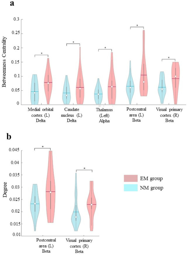
We have previously evidenced that Mindfulness Meditation (MM) in experienced meditators (EMs) is associated with long-lasting topological changes in resting state condition. However, what occurs during the meditative phase is still debated. Utilizing magnetoencephalography (MEG), the present study is aimed at comparing the topological features of the brain network in a group of EMs (n = 26) during the meditative phase with those of individuals who had no previous experience of any type of meditation (NM group, n = 29). A wide range of topological changes in the EM group as compared to the NM group has been shown. Specifically, in EMs, we have observed increased betweenness centrality in delta, alpha, and beta bands in both cortical (left medial orbital cortex, left postcentral area, and right visual primary cortex) and subcortical (left caudate nucleus and thalamus) areas. Furthermore, the degree of beta band in parietal and occipital areas of EMs was increased too. Our exploratory study suggests that the MM can change the functional brain network and provides an explanatory hypothesis on the brain circuits characterizing the meditative process.
期刊介绍:
AIMS Neuroscience is an international Open Access journal devoted to publishing peer-reviewed, high quality, original papers from all areas in the field of neuroscience. The primary focus is to provide a forum in which to expedite the speed with which theoretical neuroscience progresses toward generating testable hypotheses. In the presence of current and developing technology that offers unprecedented access to functions of the nervous system at all levels, the journal is designed to serve the role of providing the widest variety of the best theoretical views leading to suggested studies. Single blind peer review is provided for all articles and commentaries.

 求助内容:
求助内容: 应助结果提醒方式:


