Intraosseous Dermoid Presenting as an Expansile Lytic Lesion.
IF 0.7
Q4 CLINICAL NEUROLOGY
Journal of Neurological Surgery Reports
Pub Date : 2022-07-10
eCollection Date: 2022-07-01
DOI:10.1055/s-0042-1750291
引用次数: 0
Abstract
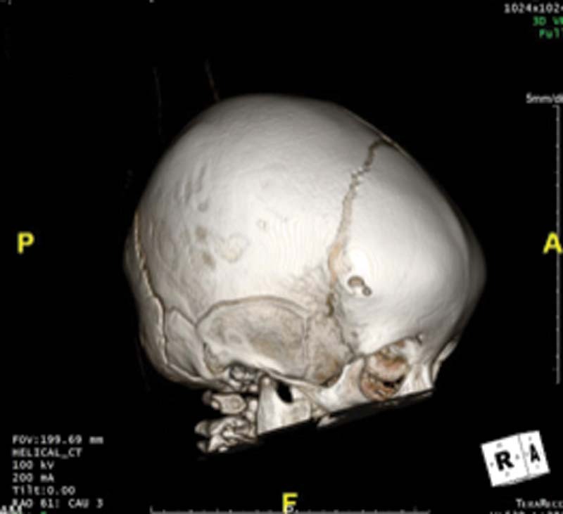
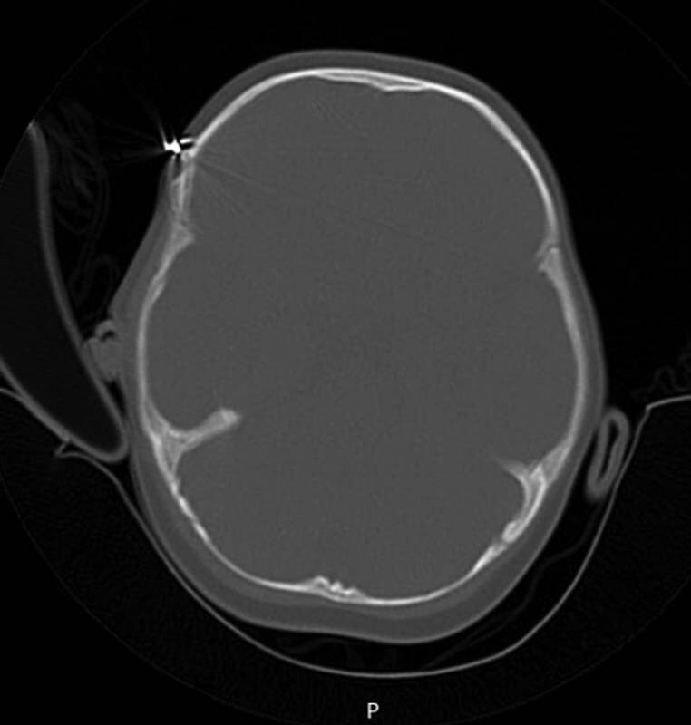
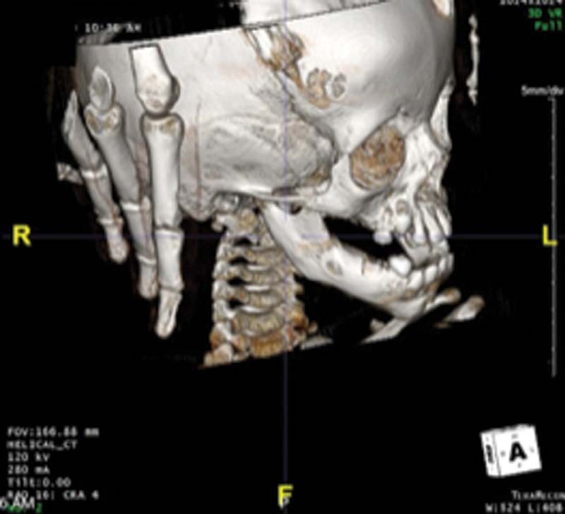
Cranial dermoids have the tendency to occur in the midline, especially near fontanelles and sutures early in the life of a patient. Here we present an unusual case of an intraosseous dermoid that presented initially as a lytic lesion, off of the midline and not associated with cranial sutures or fontanelles. The diameter of the lesion grew to approx 15 mm over time, thus the decision was made to take the child to surgery for removal of dermoid with the use of neuronavigation and cranioplasty. A dermoid cyst was confirmed on histopathologic analysis.
骨内皮样病变表现为扩张性溶解性病变。
颅皮样有发生在中线的趋势,尤其是在病人生命早期靠近囟门和缝合线的地方。我们在此报告一例不寻常的骨内皮样病变,最初表现为溶解性病变,脱离中线,与颅缝或囟门无关。随着时间的推移,病变的直径增长到大约15mm,因此我们决定带孩子进行手术,使用神经导航和颅骨成形术去除皮样。组织病理分析证实为皮样囊肿。
本文章由计算机程序翻译,如有差异,请以英文原文为准。
求助全文
约1分钟内获得全文
求助全文

 求助内容:
求助内容: 应助结果提醒方式:


