Guilherme Kuceki, Dekker C Deacon, Aaron M Secrest
{"title":"Amelanotic Melanoma Treated as Fungal Infection for Years.","authors":"Guilherme Kuceki, Dekker C Deacon, Aaron M Secrest","doi":"10.1155/2022/2598965","DOIUrl":null,"url":null,"abstract":"<p><p>This study describes a case of amelanotic lentigo maligna melanoma in a 69-year-old female that had been growing for approximately 5 years. The asymptomatic lesion had been previously diagnosed and treated as a fungal skin infection, an inflammatory rash, and an actinic keratosis that did not respond to standard treatments. Biopsy revealed confluent and nested atypical melanocytes at the dermal-epidermal junction, consistent with melanoma in situ. Excisional biopsy revealed invasive lentigo maligna melanoma, Breslow depth 0.3 mm, with positive melanoma in situ at margins. She is now 3 years post-Mohs surgery without recurrence. When working up a patient with a hypopigmented or inflammatory lesion not responding to standard therapies, physicians should always consider biopsy to rule out unusual neoplastic etiologies, such as amelanotic melanomas.</p>","PeriodicalId":9630,"journal":{"name":"Case Reports in Dermatological Medicine","volume":" ","pages":"2598965"},"PeriodicalIF":0.0000,"publicationDate":"2022-11-07","publicationTypes":"Journal Article","fieldsOfStudy":null,"isOpenAccess":false,"openAccessPdf":"https://www.ncbi.nlm.nih.gov/pmc/articles/PMC9663247/pdf/","citationCount":"1","resultStr":null,"platform":"Semanticscholar","paperid":null,"PeriodicalName":"Case Reports in Dermatological Medicine","FirstCategoryId":"1085","ListUrlMain":"https://doi.org/10.1155/2022/2598965","RegionNum":0,"RegionCategory":null,"ArticlePicture":[],"TitleCN":null,"AbstractTextCN":null,"PMCID":null,"EPubDate":"2022/1/1 0:00:00","PubModel":"eCollection","JCR":"Q3","JCRName":"Medicine","Score":null,"Total":0}
引用次数: 1
Abstract
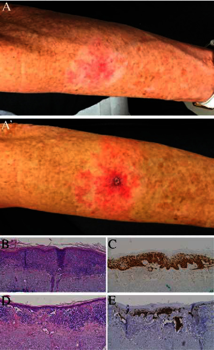
This study describes a case of amelanotic lentigo maligna melanoma in a 69-year-old female that had been growing for approximately 5 years. The asymptomatic lesion had been previously diagnosed and treated as a fungal skin infection, an inflammatory rash, and an actinic keratosis that did not respond to standard treatments. Biopsy revealed confluent and nested atypical melanocytes at the dermal-epidermal junction, consistent with melanoma in situ. Excisional biopsy revealed invasive lentigo maligna melanoma, Breslow depth 0.3 mm, with positive melanoma in situ at margins. She is now 3 years post-Mohs surgery without recurrence. When working up a patient with a hypopigmented or inflammatory lesion not responding to standard therapies, physicians should always consider biopsy to rule out unusual neoplastic etiologies, such as amelanotic melanomas.

 求助内容:
求助内容: 应助结果提醒方式:


