{"title":"Multidetector Computed Tomographic Evaluation of the Normal Characteristics of the Thymus in the Pediatric Population.","authors":"Edis Çolak, Behzat Özkan","doi":"10.5334/jbsr.2971","DOIUrl":null,"url":null,"abstract":"<p><strong>Objectives: </strong>The objective of the present study was to determine the morphologic features and measurements of the normal thymus on contrast-enhanced multi-detector computed tomography (MDCT) in subjects from the newborn period up to 18 years of age.</p><p><strong>Materials and methods: </strong>The MDCT scans obtained from 464 children with a mean age ± SD of 8.43 ± 5.60 years were retrospectively re-evaluated. The shape, margins, side predominance, density, and measurements of the thymic gland were defined for each age group.</p><p><strong>Results: </strong>A triangular thymic shape with a middle location and straight lateral contours were the most frequently seen morphologic features in children. The mean anteroposterior and transverse diameter of the thymus was 17.32 ± 4.58 and 29.99 ± 11.42 mm, respectively. The mean values for the width and thickness were 20.66 ± 5.36 and 15.15 ± 6.76 mm for the right thymic lobe, respectively; and 26.14 ± 7.85 and 14.91 ± 5.51 mm for the left, respectively. The transverse diameter of the thymus and thymic lobe dimensions decreased significantly with age, however, the anteroposterior diameter of the thymus was not significantly associated with age. Girls had higher mean thymic attenuation values compared to boys, however, this gender difference was not statistically significant (63.8 ± 22.4 HU vs. 60.1 ± 25.3, p = 0.164).</p><p><strong>Conclusion: </strong>Our study provides a better understanding of the normal thymic appearances in children that can aid in accurate diagnosis and avoid unnecessary, costly, and invasive interventions.</p>","PeriodicalId":56282,"journal":{"name":"Journal of the Belgian Society of Radiology","volume":" ","pages":"110"},"PeriodicalIF":1.3000,"publicationDate":"2022-11-17","publicationTypes":"Journal Article","fieldsOfStudy":null,"isOpenAccess":false,"openAccessPdf":"https://www.ncbi.nlm.nih.gov/pmc/articles/PMC9673598/pdf/","citationCount":"1","resultStr":null,"platform":"Semanticscholar","paperid":null,"PeriodicalName":"Journal of the Belgian Society of Radiology","FirstCategoryId":"3","ListUrlMain":"https://doi.org/10.5334/jbsr.2971","RegionNum":4,"RegionCategory":"医学","ArticlePicture":[],"TitleCN":null,"AbstractTextCN":null,"PMCID":null,"EPubDate":"2022/1/1 0:00:00","PubModel":"eCollection","JCR":"Q4","JCRName":"Medicine","Score":null,"Total":0}
引用次数: 1
Abstract
Objectives: The objective of the present study was to determine the morphologic features and measurements of the normal thymus on contrast-enhanced multi-detector computed tomography (MDCT) in subjects from the newborn period up to 18 years of age.
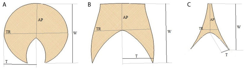
Materials and methods: The MDCT scans obtained from 464 children with a mean age ± SD of 8.43 ± 5.60 years were retrospectively re-evaluated. The shape, margins, side predominance, density, and measurements of the thymic gland were defined for each age group.
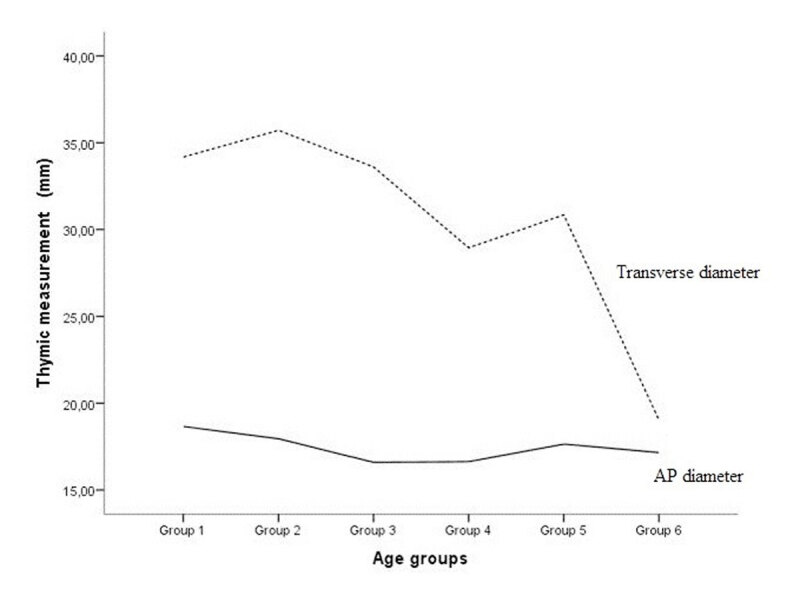
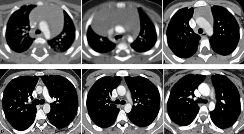
Results: A triangular thymic shape with a middle location and straight lateral contours were the most frequently seen morphologic features in children. The mean anteroposterior and transverse diameter of the thymus was 17.32 ± 4.58 and 29.99 ± 11.42 mm, respectively. The mean values for the width and thickness were 20.66 ± 5.36 and 15.15 ± 6.76 mm for the right thymic lobe, respectively; and 26.14 ± 7.85 and 14.91 ± 5.51 mm for the left, respectively. The transverse diameter of the thymus and thymic lobe dimensions decreased significantly with age, however, the anteroposterior diameter of the thymus was not significantly associated with age. Girls had higher mean thymic attenuation values compared to boys, however, this gender difference was not statistically significant (63.8 ± 22.4 HU vs. 60.1 ± 25.3, p = 0.164).
Conclusion: Our study provides a better understanding of the normal thymic appearances in children that can aid in accurate diagnosis and avoid unnecessary, costly, and invasive interventions.
期刊介绍:
The purpose of the Journal of the Belgian Society of Radiology is the publication of articles dealing with diagnostic and interventional radiology, related imaging techniques, allied sciences, and continuing education.
 求助内容:
求助内容: 应助结果提醒方式:


