Hemat Yaghoubi Mogadam, Mostafa Erfani, Mohammad Nikpassand, Masoud Mokhtary
{"title":"Evaluation of [<sup>99m</sup>Tc][Tc-HYNIC/EDDA]-Tyr as a target for metabolic tumor imaging in B16F10 melanoma tumor.","authors":"Hemat Yaghoubi Mogadam, Mostafa Erfani, Mohammad Nikpassand, Masoud Mokhtary","doi":"10.22038/AOJNMB.2021.60334.1420","DOIUrl":null,"url":null,"abstract":"<p><strong>Objectives: </strong>Clinical interest in metabolic imaging of cancer has been growing in recent years. The increase in protein metabolism of cancer cells is interesting target for metabolic tumor imaging, for which radiolabeled amino acids can be applied. The aim of this study was to evaluate a newly developed radiolabeled amino acid as an imaging protein metabolism in melanoma tumor.</p><p><strong>Methods: </strong>The radiolabeled tyrosine ([<sup>99m</sup>Tc][Tc-HYNIC/EDDA]-Tyr) was prepared and its biological properties was evaluated in B16F10 melanoma tumor. Moreover organs uptake and tumor accumulation were measured in mouse bearing B16F10 melanoma tumor.</p><p><strong>Results: </strong>Radiolabeled tyrosine was attached in B16F10 melanoma cells and showed the cell binding capacity of 13.82±0.73%. In animal study, the accumulation of radiolabeled tyrosine was observed in B16F10 melanoma tumor (2.15±0.09 %ID/g) after 30 min post injection, so that the uptake ratio of tumor to muscle was about 5.11. Through scintigraphy process the melanoma tumor clearly visualized in mice at 30 min post injection.</p><p><strong>Conclusion: </strong>These data suggest that the novel radiotracer ([<sup>99m</sup>Tc][Tc-HYNIC/EDDA]-Tyr) as an protein metabolism imaging agent, is able to transfer into melanoma cells and show great expectation for the clinical application in the imaging of melanoma tumors.</p>","PeriodicalId":8503,"journal":{"name":"Asia Oceania Journal of Nuclear Medicine and Biology","volume":"10 2","pages":"100-108"},"PeriodicalIF":0.0000,"publicationDate":"2022-01-01","publicationTypes":"Journal Article","fieldsOfStudy":null,"isOpenAccess":false,"openAccessPdf":"https://www.ncbi.nlm.nih.gov/pmc/articles/PMC9205849/pdf/","citationCount":"1","resultStr":null,"platform":"Semanticscholar","paperid":null,"PeriodicalName":"Asia Oceania Journal of Nuclear Medicine and Biology","FirstCategoryId":"1085","ListUrlMain":"https://doi.org/10.22038/AOJNMB.2021.60334.1420","RegionNum":0,"RegionCategory":null,"ArticlePicture":[],"TitleCN":null,"AbstractTextCN":null,"PMCID":null,"EPubDate":"","PubModel":"","JCR":"Q3","JCRName":"Medicine","Score":null,"Total":0}
引用次数: 1
Abstract
Objectives: Clinical interest in metabolic imaging of cancer has been growing in recent years. The increase in protein metabolism of cancer cells is interesting target for metabolic tumor imaging, for which radiolabeled amino acids can be applied. The aim of this study was to evaluate a newly developed radiolabeled amino acid as an imaging protein metabolism in melanoma tumor.
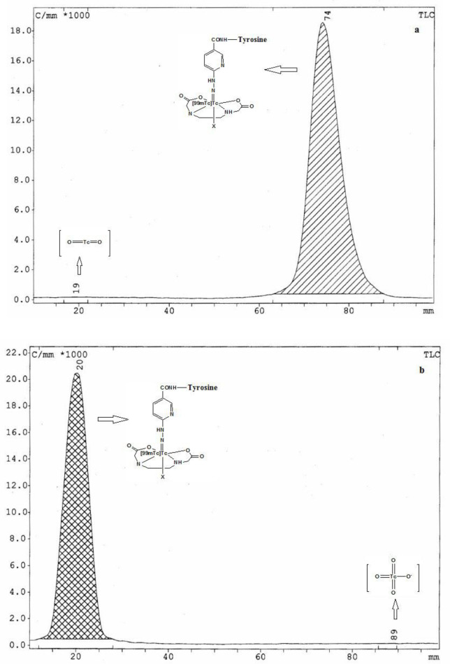
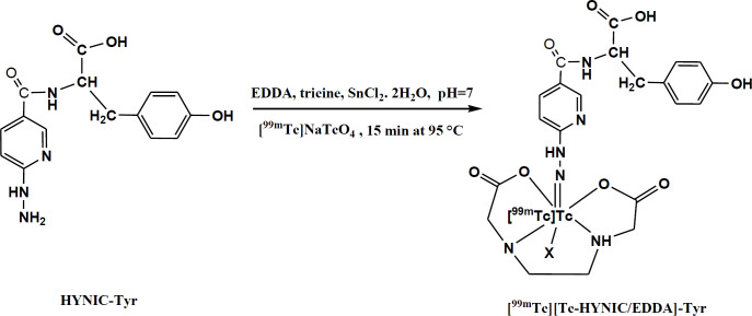
Methods: The radiolabeled tyrosine ([99mTc][Tc-HYNIC/EDDA]-Tyr) was prepared and its biological properties was evaluated in B16F10 melanoma tumor. Moreover organs uptake and tumor accumulation were measured in mouse bearing B16F10 melanoma tumor.
Results: Radiolabeled tyrosine was attached in B16F10 melanoma cells and showed the cell binding capacity of 13.82±0.73%. In animal study, the accumulation of radiolabeled tyrosine was observed in B16F10 melanoma tumor (2.15±0.09 %ID/g) after 30 min post injection, so that the uptake ratio of tumor to muscle was about 5.11. Through scintigraphy process the melanoma tumor clearly visualized in mice at 30 min post injection.
Conclusion: These data suggest that the novel radiotracer ([99mTc][Tc-HYNIC/EDDA]-Tyr) as an protein metabolism imaging agent, is able to transfer into melanoma cells and show great expectation for the clinical application in the imaging of melanoma tumors.

 求助内容:
求助内容: 应助结果提醒方式:


