Chanmi Yeon, Jeong Myo Im, Minsung Kim, Young Ro Kim, Euiheon Chung
{"title":"Cranial and Spinal Window Preparation for <i>in vivo</i> Optical Neuroimaging in Rodents and Related Experimental Techniques.","authors":"Chanmi Yeon, Jeong Myo Im, Minsung Kim, Young Ro Kim, Euiheon Chung","doi":"10.5607/en22015","DOIUrl":null,"url":null,"abstract":"<p><p>Optical neuroimaging provides an effective neuroscience tool for multi-scale investigation of the neural structures and functions, ranging from molecular, cellular activities to the inter-regional connectivity assessment. Amongst experimental preparations, the implementation of an artificial window to the central nervous system (CNS) is primarily required for optical visualization of the CNS and associated brain activities through the opaque skin and bone. Either thinning down or removing portions of the skull or spine is necessary for unobstructed long-term <i>in vivo</i> observations, for which types of the cranial and spinal window and applied materials vary depending on the study objectives. As diversely useful, a window can be designed to accommodate other experimental methods such as electrophysiology or optogenetics. Moreover, auxiliary apparatuses would allow the recording in synchrony with behavior of large-scale brain connectivity signals across the CNS, such as olfactory bulb, cerebral cortex, cerebellum, and spinal cord. Such advancements in the cranial and spinal window have resulted in a paradigm shift in neuroscience, enabling <i>in vivo</i> investigation of the brain function and dysfunction at the microscopic, cellular level. This Review addresses the types and classifications of windows used in optical neuroimaging while describing how to perform <i>in vivo</i> studies using rodent models in combination with other experimental modalities during behavioral tests. The cranial and spinal window has enabled longitudinal examination of evolving neural mechanisms via <i>in situ</i> visualization of the brain. We expect transformable and multi-functional cranial and spinal windows to become commonplace in neuroscience laboratories, further facilitating advances in optical neuroimaging systems.</p>","PeriodicalId":12263,"journal":{"name":"Experimental Neurobiology","volume":" ","pages":"131-146"},"PeriodicalIF":2.1000,"publicationDate":"2022-06-30","publicationTypes":"Journal Article","fieldsOfStudy":null,"isOpenAccess":false,"openAccessPdf":"https://ftp.ncbi.nlm.nih.gov/pub/pmc/oa_pdf/9b/a0/en-31-3-131.PMC9272117.pdf","citationCount":"1","resultStr":null,"platform":"Semanticscholar","paperid":null,"PeriodicalName":"Experimental Neurobiology","FirstCategoryId":"3","ListUrlMain":"https://doi.org/10.5607/en22015","RegionNum":4,"RegionCategory":"医学","ArticlePicture":[],"TitleCN":null,"AbstractTextCN":null,"PMCID":null,"EPubDate":"","PubModel":"","JCR":"Q3","JCRName":"MEDICINE, RESEARCH & EXPERIMENTAL","Score":null,"Total":0}
引用次数: 1
Abstract
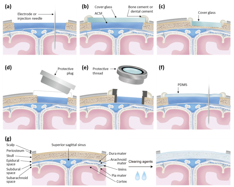
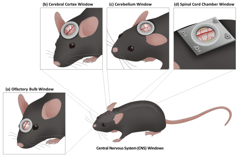
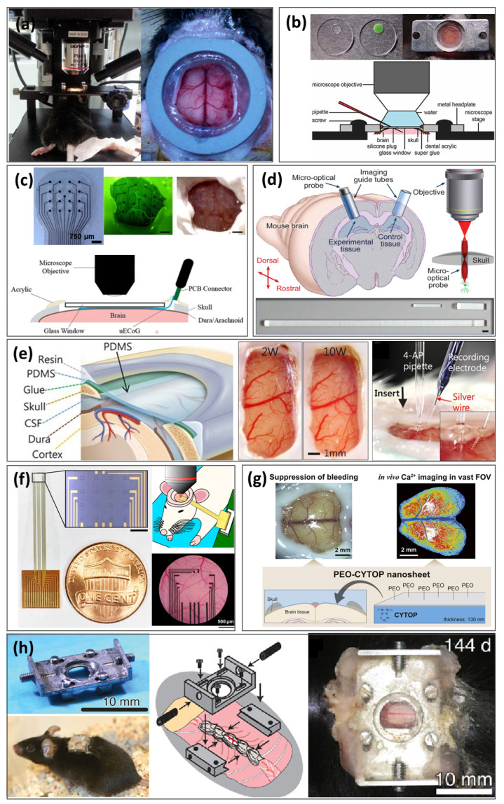
Optical neuroimaging provides an effective neuroscience tool for multi-scale investigation of the neural structures and functions, ranging from molecular, cellular activities to the inter-regional connectivity assessment. Amongst experimental preparations, the implementation of an artificial window to the central nervous system (CNS) is primarily required for optical visualization of the CNS and associated brain activities through the opaque skin and bone. Either thinning down or removing portions of the skull or spine is necessary for unobstructed long-term in vivo observations, for which types of the cranial and spinal window and applied materials vary depending on the study objectives. As diversely useful, a window can be designed to accommodate other experimental methods such as electrophysiology or optogenetics. Moreover, auxiliary apparatuses would allow the recording in synchrony with behavior of large-scale brain connectivity signals across the CNS, such as olfactory bulb, cerebral cortex, cerebellum, and spinal cord. Such advancements in the cranial and spinal window have resulted in a paradigm shift in neuroscience, enabling in vivo investigation of the brain function and dysfunction at the microscopic, cellular level. This Review addresses the types and classifications of windows used in optical neuroimaging while describing how to perform in vivo studies using rodent models in combination with other experimental modalities during behavioral tests. The cranial and spinal window has enabled longitudinal examination of evolving neural mechanisms via in situ visualization of the brain. We expect transformable and multi-functional cranial and spinal windows to become commonplace in neuroscience laboratories, further facilitating advances in optical neuroimaging systems.
期刊介绍:
Experimental Neurobiology is an international forum for interdisciplinary investigations of the nervous system. The journal aims to publish papers that present novel observations in all fields of neuroscience, encompassing cellular & molecular neuroscience, development/differentiation/plasticity, neurobiology of disease, systems/cognitive/behavioral neuroscience, drug development & industrial application, brain-machine interface, methodologies/tools, and clinical neuroscience. It should be of interest to a broad scientific audience working on the biochemical, molecular biological, cell biological, pharmacological, physiological, psychophysical, clinical, anatomical, cognitive, and biotechnological aspects of neuroscience. The journal publishes both original research articles and review articles. Experimental Neurobiology is an open access, peer-reviewed online journal. The journal is published jointly by The Korean Society for Brain and Neural Sciences & The Korean Society for Neurodegenerative Disease.
 求助内容:
求助内容: 应助结果提醒方式:


