Natarajan Ramya, Murhekar Kanchan, Raja Anand, Sundersingh Shirley
{"title":"A Case Report: Mucinous Tubular and Spindle Cell Carcinoma of Kidney with Spindle Cell Predominance Mimicking Mesenchymal Tumour.","authors":"Natarajan Ramya, Murhekar Kanchan, Raja Anand, Sundersingh Shirley","doi":"10.15586/jkcvhl.v9i4.203","DOIUrl":null,"url":null,"abstract":"<p><p>Mucinous tubular and spindle cell carcinoma (MTSCC) of kidney is a rare variant of renal cell carcinoma which was first described in the 2004 World Health Organization classification of tumours of the kidney. Morphologically, MTSCC is composed of tubules merging with bland-appearing spindle cells in a myxoid/mucinous stroma. Diverse morphological patterns have been reported in MTSCC; however, a spindle cell predominant morphology mimicking a mesenchymal tumour is rare and only two cases have been reported so far. We report a case of MTSCC with spindle cell predominance in kidney which was a diagnostic challenge. Though MTSCC usually shows an indolent course, there have been cases showing aggressive behaviour even with bland morphology. Hence, a thorough histopathological evaluation with ancillary studies are required to differentiate spindle cell predominant MTSCC from its mimics. Our case was a 40-year-old female who was incidentally found to have a well-defined hypodense lesion measuring around 2 cm in the upper pole of the right kidney. Right partial nephrectomy was performed which showed a 2.7 × 2.5 × 2 cm well-defined grey tan tumour without necrosis or haemorrhage, limited to kidney. Histopathological examination showed sheets of bland-appearing spindle cells mimicking a mesenchymal tumour. The tumour was extensively sampled, revealing small foci of tubule formation and mucinous stroma. Tumour cells were positive for CK7, AMACR, and PAX8. A final diagnosis of MTSCC was made. Hereby, we discuss ways of differentiating MTSCC from other spindle cell tumours of the kidney.</p>","PeriodicalId":44291,"journal":{"name":"Journal of Kidney Cancer and VHL","volume":" ","pages":"10-13"},"PeriodicalIF":1.9000,"publicationDate":"2022-10-31","publicationTypes":"Journal Article","fieldsOfStudy":null,"isOpenAccess":false,"openAccessPdf":"https://www.ncbi.nlm.nih.gov/pmc/articles/PMC9634216/pdf/","citationCount":"1","resultStr":null,"platform":"Semanticscholar","paperid":null,"PeriodicalName":"Journal of Kidney Cancer and VHL","FirstCategoryId":"1085","ListUrlMain":"https://doi.org/10.15586/jkcvhl.v9i4.203","RegionNum":0,"RegionCategory":null,"ArticlePicture":[],"TitleCN":null,"AbstractTextCN":null,"PMCID":null,"EPubDate":"2022/1/1 0:00:00","PubModel":"eCollection","JCR":"Q3","JCRName":"ONCOLOGY","Score":null,"Total":0}
引用次数: 1
Abstract
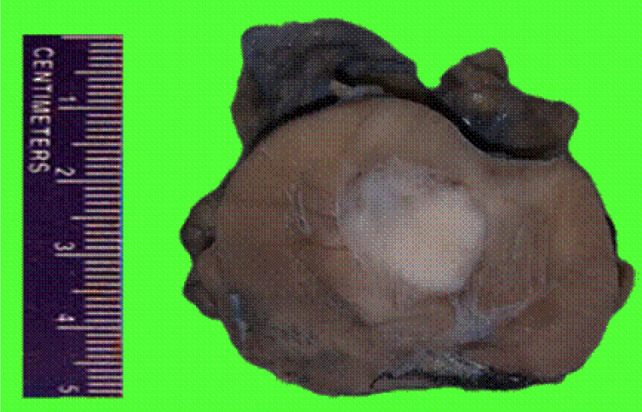
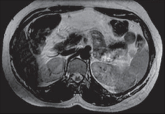
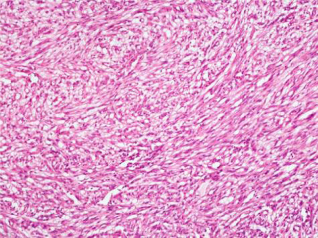
Mucinous tubular and spindle cell carcinoma (MTSCC) of kidney is a rare variant of renal cell carcinoma which was first described in the 2004 World Health Organization classification of tumours of the kidney. Morphologically, MTSCC is composed of tubules merging with bland-appearing spindle cells in a myxoid/mucinous stroma. Diverse morphological patterns have been reported in MTSCC; however, a spindle cell predominant morphology mimicking a mesenchymal tumour is rare and only two cases have been reported so far. We report a case of MTSCC with spindle cell predominance in kidney which was a diagnostic challenge. Though MTSCC usually shows an indolent course, there have been cases showing aggressive behaviour even with bland morphology. Hence, a thorough histopathological evaluation with ancillary studies are required to differentiate spindle cell predominant MTSCC from its mimics. Our case was a 40-year-old female who was incidentally found to have a well-defined hypodense lesion measuring around 2 cm in the upper pole of the right kidney. Right partial nephrectomy was performed which showed a 2.7 × 2.5 × 2 cm well-defined grey tan tumour without necrosis or haemorrhage, limited to kidney. Histopathological examination showed sheets of bland-appearing spindle cells mimicking a mesenchymal tumour. The tumour was extensively sampled, revealing small foci of tubule formation and mucinous stroma. Tumour cells were positive for CK7, AMACR, and PAX8. A final diagnosis of MTSCC was made. Hereby, we discuss ways of differentiating MTSCC from other spindle cell tumours of the kidney.

 求助内容:
求助内容: 应助结果提醒方式:


