Myocardium Assessment by Relaxation along Fictitious Field, Extracellular Volume, Feature Tracking, and Myocardial Strain in Hypertensive Patients with Left Ventricular Hypertrophy.
Seyed Amir Mirmojarabian, Eveliina Lammentausta, Esa Liukkonen, Lauri Ahvenjärvi, Juhani Junttila, Miika T Nieminen, Timo Liimatainen
{"title":"Myocardium Assessment by Relaxation along Fictitious Field, Extracellular Volume, Feature Tracking, and Myocardial Strain in Hypertensive Patients with Left Ventricular Hypertrophy.","authors":"Seyed Amir Mirmojarabian, Eveliina Lammentausta, Esa Liukkonen, Lauri Ahvenjärvi, Juhani Junttila, Miika T Nieminen, Timo Liimatainen","doi":"10.1155/2022/9198691","DOIUrl":null,"url":null,"abstract":"<p><strong>Background: </strong>Previous research has shown impaired global longitudinal strain (GLS) and slightly elevated extracellular volume fraction (ECV) in hypertensive patients with left ventricular hypertrophy (HTN LVH). Up to now, only little attention has been paid to interactions between macromolecules and free water in hypertrophied myocardium.</p><p><strong>Purpose: </strong>To evaluate the feasibility of relaxation along a fictitious field with rank 2 (RAFF2) in HTN LVH patients. <i>Study Type.</i> Single institutional case control.</p><p><strong>Subjects: </strong>9 HTN LVH (age, 69 ± 10 years) and 11 control subjects (age, 54 ± 12 years). <i>Field Strength/Sequence.</i> Relaxation time mapping (<i>T</i> <sub>1</sub>, <i>T</i> <sub>1<i>ρ</i></sub> , and <i>T</i> <sub>RAFF2</sub> with 11.8 <i>μ</i>T maximum radio frequency field amplitude) was performed at 1.5 T using a Siemens Aera (Erlangen, Germany) scanner equipped with an 18-channel body array coil. <i>Assessment.</i> ECV was calculated using pre- and postcontrast <i>T</i> <sub>1</sub>, and global strains parameters were assessed by Segment CMR (Medviso AB Co, Sweden). The parametric maps of <i>T</i> <sub>1<i>ρ</i></sub> and <i>T</i> <sub>RAFF2</sub> were computed using a monoexponential model, while the Bloch-McConnell equations were solved numerically to model effect of the chemical exchange during radio frequency pulses. <i>Statistical Tests.</i> Parametric maps were averaged over myocardium for each subject to be used in statistical analysis. Kolmogorov-Smirnov was used as the normality test followed by Student's t-test and Pearson's correlation to determine the difference between the HTN LVH patients and controls along with Hedges' <i>g</i> effect size and the association between variables, respectively.</p><p><strong>Results: </strong><i>T</i> <sub>RAFF2</sub> decreased statistically (83 ± 2 ms vs 88 ± 6 ms, <i>P</i> < 0.031), and global longitudinal strain was impaired (GLS, -14 ± 3 vs - 18 ± 2, <i>P</i> < 0.002) in HTN LVH patients compared to the controls, respectively. Also, significant negative correlation was found between <i>T</i> <sub>RAFF2</sub> and GLS (<i>r</i> = -0.53, <i>P</i> < 0.05). <i>Data Conclusion.</i> Our results suggest that <i>T</i> <sub>RAFF2</sub> decrease in HTN LVH patients may be explained by gradual collagen accumulation which can be reflected in GLS changes. Most likely, it increases the water proton interactions and consequently decreases <i>T</i> <sub>RAFF2</sub> before myocardial scarring.</p>","PeriodicalId":47063,"journal":{"name":"International Journal of Biomedical Imaging","volume":null,"pages":null},"PeriodicalIF":3.3000,"publicationDate":"2022-06-23","publicationTypes":"Journal Article","fieldsOfStudy":null,"isOpenAccess":false,"openAccessPdf":"https://www.ncbi.nlm.nih.gov/pmc/articles/PMC9246602/pdf/","citationCount":"0","resultStr":null,"platform":"Semanticscholar","paperid":null,"PeriodicalName":"International Journal of Biomedical Imaging","FirstCategoryId":"1085","ListUrlMain":"https://doi.org/10.1155/2022/9198691","RegionNum":0,"RegionCategory":null,"ArticlePicture":[],"TitleCN":null,"AbstractTextCN":null,"PMCID":null,"EPubDate":"2022/1/1 0:00:00","PubModel":"eCollection","JCR":"Q2","JCRName":"ENGINEERING, BIOMEDICAL","Score":null,"Total":0}
引用次数: 0
Abstract
Background: Previous research has shown impaired global longitudinal strain (GLS) and slightly elevated extracellular volume fraction (ECV) in hypertensive patients with left ventricular hypertrophy (HTN LVH). Up to now, only little attention has been paid to interactions between macromolecules and free water in hypertrophied myocardium.
Purpose: To evaluate the feasibility of relaxation along a fictitious field with rank 2 (RAFF2) in HTN LVH patients. Study Type. Single institutional case control.
Subjects: 9 HTN LVH (age, 69 ± 10 years) and 11 control subjects (age, 54 ± 12 years). Field Strength/Sequence. Relaxation time mapping (T1, T1ρ , and TRAFF2 with 11.8 μT maximum radio frequency field amplitude) was performed at 1.5 T using a Siemens Aera (Erlangen, Germany) scanner equipped with an 18-channel body array coil. Assessment. ECV was calculated using pre- and postcontrast T1, and global strains parameters were assessed by Segment CMR (Medviso AB Co, Sweden). The parametric maps of T1ρ and TRAFF2 were computed using a monoexponential model, while the Bloch-McConnell equations were solved numerically to model effect of the chemical exchange during radio frequency pulses. Statistical Tests. Parametric maps were averaged over myocardium for each subject to be used in statistical analysis. Kolmogorov-Smirnov was used as the normality test followed by Student's t-test and Pearson's correlation to determine the difference between the HTN LVH patients and controls along with Hedges' g effect size and the association between variables, respectively.
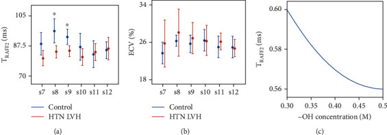
Results: TRAFF2 decreased statistically (83 ± 2 ms vs 88 ± 6 ms, P < 0.031), and global longitudinal strain was impaired (GLS, -14 ± 3 vs - 18 ± 2, P < 0.002) in HTN LVH patients compared to the controls, respectively. Also, significant negative correlation was found between TRAFF2 and GLS (r = -0.53, P < 0.05). Data Conclusion. Our results suggest that TRAFF2 decrease in HTN LVH patients may be explained by gradual collagen accumulation which can be reflected in GLS changes. Most likely, it increases the water proton interactions and consequently decreases TRAFF2 before myocardial scarring.
期刊介绍:
The International Journal of Biomedical Imaging is managed by a board of editors comprising internationally renowned active researchers. The journal is freely accessible online and also offered for purchase in print format. It employs a web-based review system to ensure swift turnaround times while maintaining high standards. In addition to regular issues, special issues are organized by guest editors. The subject areas covered include (but are not limited to):
Digital radiography and tomosynthesis
X-ray computed tomography (CT)
Magnetic resonance imaging (MRI)
Single photon emission computed tomography (SPECT)
Positron emission tomography (PET)
Ultrasound imaging
Diffuse optical tomography, coherence, fluorescence, bioluminescence tomography, impedance tomography
Neutron imaging for biomedical applications
Magnetic and optical spectroscopy, and optical biopsy
Optical, electron, scanning tunneling/atomic force microscopy
Small animal imaging
Functional, cellular, and molecular imaging
Imaging assays for screening and molecular analysis
Microarray image analysis and bioinformatics
Emerging biomedical imaging techniques
Imaging modality fusion
Biomedical imaging instrumentation
Biomedical image processing, pattern recognition, and analysis
Biomedical image visualization, compression, transmission, and storage
Imaging and modeling related to systems biology and systems biomedicine
Applied mathematics, applied physics, and chemistry related to biomedical imaging
Grid-enabling technology for biomedical imaging and informatics

 求助内容:
求助内容: 应助结果提醒方式:


