Insidious Onset of Pulmonary Langerhans Cell Histiocytosis During Oncological Follow-Up.

IF 2 4区 医学 Q4 Medicine
Journal of the Belgian Society of Radiology Pub Date : 2022-10-20 eCollection Date: 2022-01-01 DOI:10.5334/jbsr.2913
Nico Hustings, Eveline Slachmuylders, Adriana Dubbeldam
{"title":"Insidious Onset of Pulmonary Langerhans Cell Histiocytosis During Oncological Follow-Up.","authors":"Nico Hustings,&nbsp;Eveline Slachmuylders,&nbsp;Adriana Dubbeldam","doi":"10.5334/jbsr.2913","DOIUrl":null,"url":null,"abstract":"<p><p><b>Teaching Point:</b> Pulmonary Langerhans cell histiocytosis (PLCH) has to be included in the differential diagnosis of cystic pulmonary lesions on chest computed tomography (CT). CT has important diagnostic value by demonstrating initial centrilobular nodules that in time cavitate and transform into cysts, typically sparing the costophrenic angles.</p>","PeriodicalId":56282,"journal":{"name":"Journal of the Belgian Society of Radiology","volume":null,"pages":null},"PeriodicalIF":2.0000,"publicationDate":"2022-10-20","publicationTypes":"Journal Article","fieldsOfStudy":null,"isOpenAccess":false,"openAccessPdf":"https://www.ncbi.nlm.nih.gov/pmc/articles/PMC9585974/pdf/","citationCount":"0","resultStr":null,"platform":"Semanticscholar","paperid":null,"PeriodicalName":"Journal of the Belgian Society of Radiology","FirstCategoryId":"3","ListUrlMain":"https://doi.org/10.5334/jbsr.2913","RegionNum":4,"RegionCategory":"医学","ArticlePicture":[],"TitleCN":null,"AbstractTextCN":null,"PMCID":null,"EPubDate":"2022/1/1 0:00:00","PubModel":"eCollection","JCR":"Q4","JCRName":"Medicine","Score":null,"Total":0}
引用次数: 0

Abstract

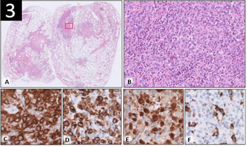
Teaching Point: Pulmonary Langerhans cell histiocytosis (PLCH) has to be included in the differential diagnosis of cystic pulmonary lesions on chest computed tomography (CT). CT has important diagnostic value by demonstrating initial centrilobular nodules that in time cavitate and transform into cysts, typically sparing the costophrenic angles.

Abstract Image

Abstract Image

Abstract Image

肿瘤随访期间肺朗格汉斯细胞组织细胞增多症的隐匿发病。
教学要点:肺朗格汉斯细胞组织细胞增多症(PLCH)必须包括在胸部计算机断层扫描(CT)的囊性肺病变鉴别诊断中。CT具有重要的诊断价值,它能及时显示小叶中心结节空化并转变为囊肿,通常不影响肋膈角。
本文章由计算机程序翻译,如有差异,请以英文原文为准。
求助全文
约1分钟内获得全文 求助全文
来源期刊
Journal of the Belgian Society of Radiology
Journal of the Belgian Society of Radiology Medicine-Radiology, Nuclear Medicine and Imaging
CiteScore
0.60
自引率
5.00%
发文量
0
审稿时长
6-12 weeks
期刊介绍: The purpose of the Journal of the Belgian Society of Radiology is the publication of articles dealing with diagnostic and interventional radiology, related imaging techniques, allied sciences, and continuing education.
×
引用
GB/T 7714-2015
复制
MLA
复制
APA
复制
导出至
BibTeX EndNote RefMan NoteFirst NoteExpress
×
提示
您的信息不完整,为了账户安全,请先补充。
现在去补充
×
提示
您因"违规操作"
具体请查看互助需知
我知道了
×
提示
确定
请完成安全验证×
copy
已复制链接
快去分享给好友吧!
我知道了
右上角分享
点击右上角分享
0
联系我们:info@booksci.cn Book学术提供免费学术资源搜索服务,方便国内外学者检索中英文文献。致力于提供最便捷和优质的服务体验。 Copyright © 2023 布克学术 All rights reserved.
京ICP备2023020795号-1
ghs 京公网安备 11010802042870号
Book学术文献互助
Book学术文献互助群
群 号:481959085
Book学术官方微信