{"title":"Atypical Hepatic Hemangioma with Fluid-Fluid Level on CT and MRI: Emphasis on Added Value of Contrast-Enhanced Ultrasound Findings.","authors":"Ye Rin Kim, Ji Eun Lee, Min Jung Jung","doi":"10.5334/jbsr.2824","DOIUrl":null,"url":null,"abstract":"<p><p>We present an atypical case of a patient with hepatic hemangiomas showing fluid-fluid levels on computed tomography (CT) and magnetic resonance imaging (MRI). None of the lesions showed contrast enhancement, mimicking complicated hepatic cysts or metastasis with hemorrhagic content. On contrast-enhanced ultrasound the lesions showed peripheral nodular enhancement with complete fill-in on late phases, suggestive of hepatic hemangioma. <b>Teaching point:</b> Contrast-enhanced ultrasound (CE-US) may be useful in diagnosing atypical hepatic hemangioma showing fluid-fluid levels on computed tomography (CT) or magnetic resonance imaging (MRI).</p>","PeriodicalId":56282,"journal":{"name":"Journal of the Belgian Society of Radiology","volume":" ","pages":"56"},"PeriodicalIF":1.3000,"publicationDate":"2022-06-09","publicationTypes":"Journal Article","fieldsOfStudy":null,"isOpenAccess":false,"openAccessPdf":"https://www.ncbi.nlm.nih.gov/pmc/articles/PMC9187241/pdf/","citationCount":"0","resultStr":null,"platform":"Semanticscholar","paperid":null,"PeriodicalName":"Journal of the Belgian Society of Radiology","FirstCategoryId":"3","ListUrlMain":"https://doi.org/10.5334/jbsr.2824","RegionNum":4,"RegionCategory":"医学","ArticlePicture":[],"TitleCN":null,"AbstractTextCN":null,"PMCID":null,"EPubDate":"2022/1/1 0:00:00","PubModel":"eCollection","JCR":"Q4","JCRName":"Medicine","Score":null,"Total":0}
引用次数: 0
Abstract
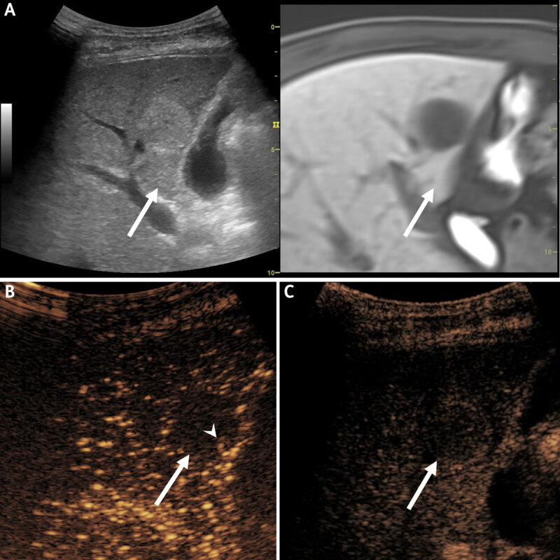
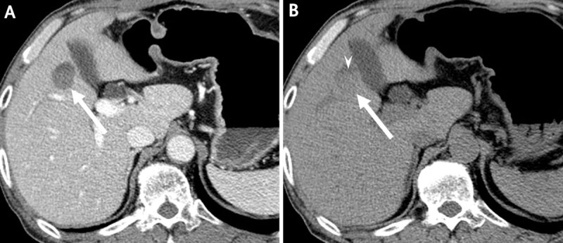
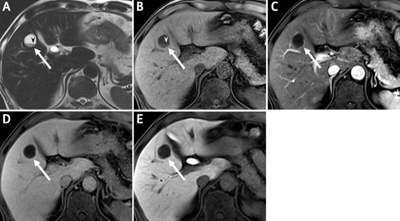
We present an atypical case of a patient with hepatic hemangiomas showing fluid-fluid levels on computed tomography (CT) and magnetic resonance imaging (MRI). None of the lesions showed contrast enhancement, mimicking complicated hepatic cysts or metastasis with hemorrhagic content. On contrast-enhanced ultrasound the lesions showed peripheral nodular enhancement with complete fill-in on late phases, suggestive of hepatic hemangioma. Teaching point: Contrast-enhanced ultrasound (CE-US) may be useful in diagnosing atypical hepatic hemangioma showing fluid-fluid levels on computed tomography (CT) or magnetic resonance imaging (MRI).
期刊介绍:
The purpose of the Journal of the Belgian Society of Radiology is the publication of articles dealing with diagnostic and interventional radiology, related imaging techniques, allied sciences, and continuing education.
 求助内容:
求助内容: 应助结果提醒方式:


