Lopez Dominguez Johanny, Olayemi Sokumbi, Misty M Hobbs, Liuyan Jiang
{"title":"Polarization of Macrophages in Granulomatous Cutaneous T Cell Lymphoma Granulomatous Mycosis Fungoides Microenvironment.","authors":"Lopez Dominguez Johanny, Olayemi Sokumbi, Misty M Hobbs, Liuyan Jiang","doi":"10.3390/dermatopathology9010009","DOIUrl":null,"url":null,"abstract":"<p><p>Polarization of tumor associated macrophages (TAMs) has been shown to have prognostic significance in different cancer types. This study evaluates the macrophage subtypes that predominates in GMF. Cases of GCTCL from 2007-2020 were identified (<i>n</i> = 6), clinical data was extracted from the electronic medical record, and all pathology slides were reviewed to confirm the diagnosis. Immunohistochemistry (IHC) studies were performed to characterize M1 and M2 macrophage polarization. CD68 (PGM1), pSTAT1, and CD163 were used as pan macrophage, M1, and M2 markers, respectively. The macrophages with positive staining at hot spot per high power field were counted and recorded for data analysis. The average age of patients was 60.5 years [range, 21-78], five patients (83%) were women and 1 (17%) was a man. Five patients were Caucasian (83%), and 1 was Black/African American (17%). Two patients had late stage GMF with M2 (CD163) predominance and the other three had early stage GMF with M1 (pSTAT1) predominance. Our study suggests that macrophage polarization present in GMF tends to be M1 in early stages and M2 in advanced stages. Additional studies are needed to further elucidate the microenvironment of macrophages present in GMF. Such findings may lead to prognostic and therapeutic advances in GMF.</p>","PeriodicalId":42885,"journal":{"name":"Dermatopathology","volume":" ","pages":"54-59"},"PeriodicalIF":1.7000,"publicationDate":"2022-02-25","publicationTypes":"Journal Article","fieldsOfStudy":null,"isOpenAccess":false,"openAccessPdf":"https://www.ncbi.nlm.nih.gov/pmc/articles/PMC8946979/pdf/","citationCount":"0","resultStr":null,"platform":"Semanticscholar","paperid":null,"PeriodicalName":"Dermatopathology","FirstCategoryId":"1085","ListUrlMain":"https://doi.org/10.3390/dermatopathology9010009","RegionNum":0,"RegionCategory":null,"ArticlePicture":[],"TitleCN":null,"AbstractTextCN":null,"PMCID":null,"EPubDate":"","PubModel":"","JCR":"Q3","JCRName":"DERMATOLOGY","Score":null,"Total":0}
引用次数: 0
Abstract
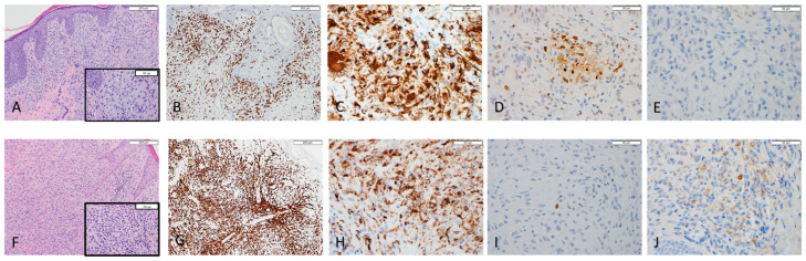
Polarization of tumor associated macrophages (TAMs) has been shown to have prognostic significance in different cancer types. This study evaluates the macrophage subtypes that predominates in GMF. Cases of GCTCL from 2007-2020 were identified (n = 6), clinical data was extracted from the electronic medical record, and all pathology slides were reviewed to confirm the diagnosis. Immunohistochemistry (IHC) studies were performed to characterize M1 and M2 macrophage polarization. CD68 (PGM1), pSTAT1, and CD163 were used as pan macrophage, M1, and M2 markers, respectively. The macrophages with positive staining at hot spot per high power field were counted and recorded for data analysis. The average age of patients was 60.5 years [range, 21-78], five patients (83%) were women and 1 (17%) was a man. Five patients were Caucasian (83%), and 1 was Black/African American (17%). Two patients had late stage GMF with M2 (CD163) predominance and the other three had early stage GMF with M1 (pSTAT1) predominance. Our study suggests that macrophage polarization present in GMF tends to be M1 in early stages and M2 in advanced stages. Additional studies are needed to further elucidate the microenvironment of macrophages present in GMF. Such findings may lead to prognostic and therapeutic advances in GMF.

 求助内容:
求助内容: 应助结果提醒方式:


