Juan Fernando Ortiz, Jennifer M Argudo, Mario Yépez, Juan Andrés Moncayo, Hyder Tamton, Alex S Aguirre, Ghanshyam Patel, Meghdeep Sen, Ayushi Mistry, Ray Yuen, Ahmed Eissa-Garces, Diego Ojeda, Samir Ruxmohan
{"title":"Neuroimaging in the Rare Sleep Disorder of Kleine-Levin Syndrome: A Systematic Review.","authors":"Juan Fernando Ortiz, Jennifer M Argudo, Mario Yépez, Juan Andrés Moncayo, Hyder Tamton, Alex S Aguirre, Ghanshyam Patel, Meghdeep Sen, Ayushi Mistry, Ray Yuen, Ahmed Eissa-Garces, Diego Ojeda, Samir Ruxmohan","doi":"10.3390/clockssleep4020025","DOIUrl":null,"url":null,"abstract":"<p><p>Kleine-Levin syndrome (KLS) is characterized by episodes of hypersomnia. Additionally, these patients can present with hyperphagia, hypersexuality, abnormal behavior, and cognitive dysfunction. Functional neuroimaging studies such as fMRI-BOLD, Positron Emission Tomography (PET) or SPECT help us understand the neuropathological bases of different disorders. We conducted a systematic review to investigate the neuroimaging features of KLS patients and their clinical correlations. This systematic review was conducted by following the Meta-Analysis of Observational Studies in Epidemiology (MOOSE) and PRISMA protocol reporting guidelines. We aim to investigate the clinical correlation with neuroimaging among patients with KLS. We included only studies written in the English language in the last 20 years, conducted on humans; 10 studies were included. We excluded systematic reviews, metanalysis, and case reports. We found that there are changes in functional imaging studies during the symptomatic and asymptomatic periods as well as in between episodes in patients with K.L.S. The areas most reported as affected were the hypothalamic and thalamic regions, which showed hypoperfusion and, in a few cases, hyperperfusion; areas such as the frontal, parietal, occipital and the prefrontal cortex all showed alterations in cerebral perfusion. These changes in cerebral blood flow and regions vary according to the imaging (SPECT, PET SCAN, or fMRI) and the task performed while imaging was performed. We encountered conflicting data between studies. Hyper insomnia, the main feature of this disease during the symptomatic periods, was associated with decreased thalamic activity. Other features of K.L.S., such as apathy, hypersexuality, and depersonalization, were also correlated with functional imaging changes. There were also findings that correlated with working memory deficits seen in this stage during the asymptomatic periods. Hyperactivity of the thalamus and hypothalamus were the main features shown during the asymptomatic period. Additionally, functional imaging tends to improve with a longer course of the disease, which suggests that K.L.S. patients outgrow the disease. These findings should caution physicians when analyzing and correlating neuroimaging findings with the disease.</p>","PeriodicalId":33568,"journal":{"name":"Clocks & Sleep","volume":null,"pages":null},"PeriodicalIF":2.1000,"publicationDate":"2022-05-31","publicationTypes":"Journal Article","fieldsOfStudy":null,"isOpenAccess":false,"openAccessPdf":"https://www.ncbi.nlm.nih.gov/pmc/articles/PMC9221874/pdf/","citationCount":"0","resultStr":null,"platform":"Semanticscholar","paperid":null,"PeriodicalName":"Clocks & Sleep","FirstCategoryId":"1085","ListUrlMain":"https://doi.org/10.3390/clockssleep4020025","RegionNum":0,"RegionCategory":null,"ArticlePicture":[],"TitleCN":null,"AbstractTextCN":null,"PMCID":null,"EPubDate":"","PubModel":"","JCR":"Q3","JCRName":"CLINICAL NEUROLOGY","Score":null,"Total":0}
引用次数: 0
Abstract
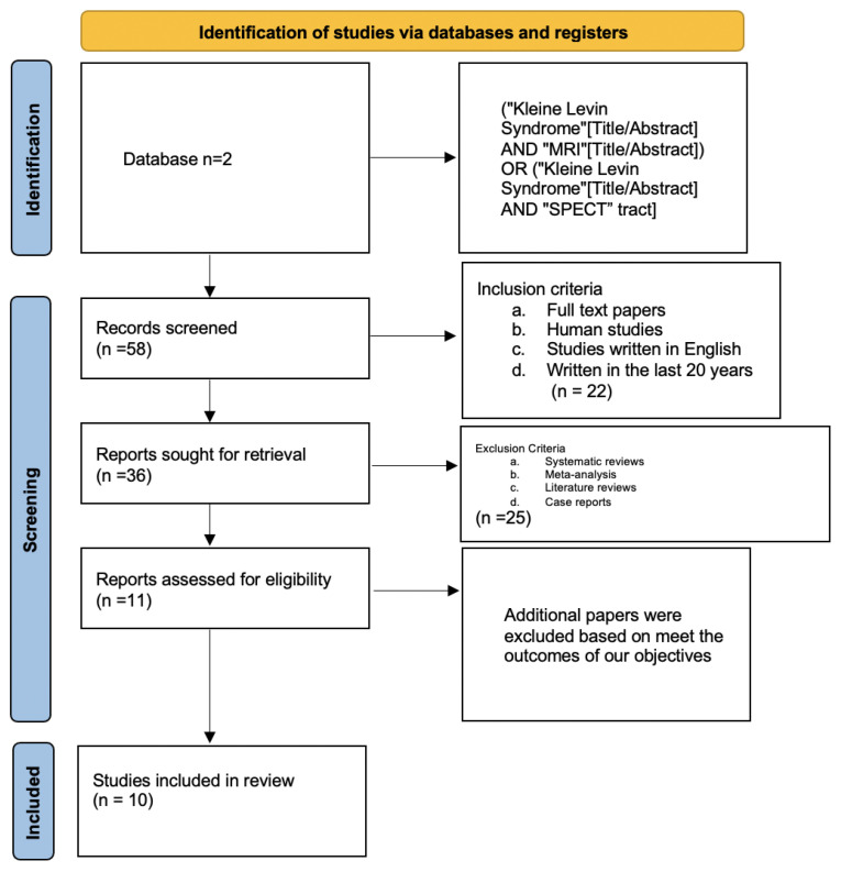
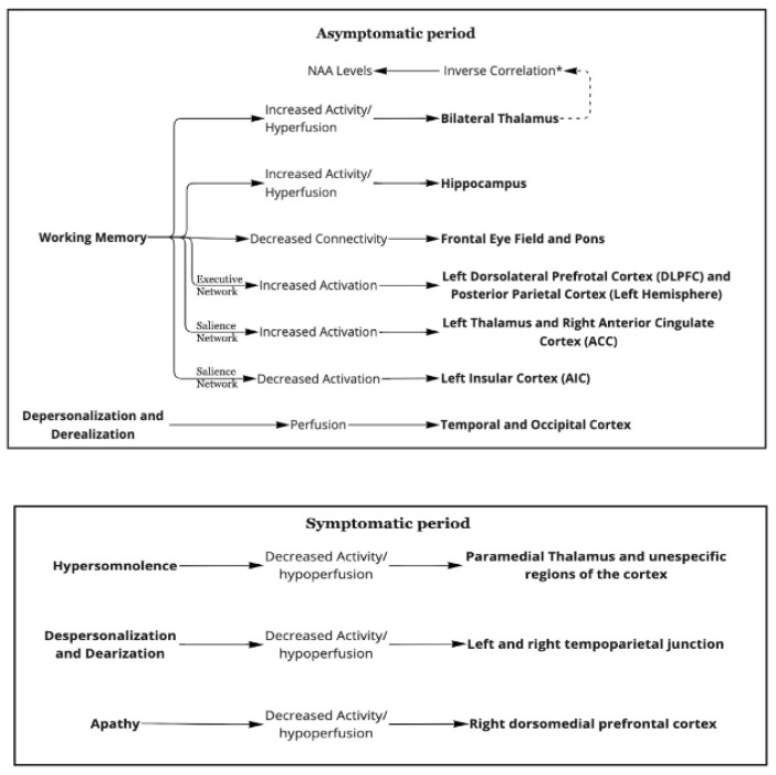
Kleine-Levin syndrome (KLS) is characterized by episodes of hypersomnia. Additionally, these patients can present with hyperphagia, hypersexuality, abnormal behavior, and cognitive dysfunction. Functional neuroimaging studies such as fMRI-BOLD, Positron Emission Tomography (PET) or SPECT help us understand the neuropathological bases of different disorders. We conducted a systematic review to investigate the neuroimaging features of KLS patients and their clinical correlations. This systematic review was conducted by following the Meta-Analysis of Observational Studies in Epidemiology (MOOSE) and PRISMA protocol reporting guidelines. We aim to investigate the clinical correlation with neuroimaging among patients with KLS. We included only studies written in the English language in the last 20 years, conducted on humans; 10 studies were included. We excluded systematic reviews, metanalysis, and case reports. We found that there are changes in functional imaging studies during the symptomatic and asymptomatic periods as well as in between episodes in patients with K.L.S. The areas most reported as affected were the hypothalamic and thalamic regions, which showed hypoperfusion and, in a few cases, hyperperfusion; areas such as the frontal, parietal, occipital and the prefrontal cortex all showed alterations in cerebral perfusion. These changes in cerebral blood flow and regions vary according to the imaging (SPECT, PET SCAN, or fMRI) and the task performed while imaging was performed. We encountered conflicting data between studies. Hyper insomnia, the main feature of this disease during the symptomatic periods, was associated with decreased thalamic activity. Other features of K.L.S., such as apathy, hypersexuality, and depersonalization, were also correlated with functional imaging changes. There were also findings that correlated with working memory deficits seen in this stage during the asymptomatic periods. Hyperactivity of the thalamus and hypothalamus were the main features shown during the asymptomatic period. Additionally, functional imaging tends to improve with a longer course of the disease, which suggests that K.L.S. patients outgrow the disease. These findings should caution physicians when analyzing and correlating neuroimaging findings with the disease.

 求助内容:
求助内容: 应助结果提醒方式:


