{"title":"Clinicopathological assessment of cancer/testis antigens NY‑ESO‑1 and MAGE‑A4 in osteosarcoma.","authors":"Kazuhiko Hashimoto, Shunji Nishimura, Tomohiko Ito, Naohiro Oka, Ryosuke Kakinoki, Masao Akagi","doi":"10.4081/ejh.2022.3377","DOIUrl":null,"url":null,"abstract":"<p><p>The cancer/testis antigens (CTAs), New York esophageal squamous cell carcinoma-1 (NY-ESO-1) and melanoma antigen gene (MAGE)-A4 are normally restricted to male germ cells but are aberrantly expressed in several cancers. Considering the limited information regarding their significance in osteosarcoma (OS), the purpose of this study was to determine the clinical significance of NY-ESO-1 and MAGE-A4 expression in OS. Nine patients with OS treated at Kindai University Hospital were included in the study. The median age was 27 years, and median follow-up period was 40 months. The specimens obtained at the time of biopsy were used to perform immunostaining for NY-ESO, MAGE-A4, p53, and Ki-67. The positive cell rates and positive case rates of NY-ESO, MAGE-A4, p53, and Ki-67 were calculated. The correlation between the positive cell rate of immunohistochemical markers was also calculated. The correlation between the positive cell rate of NY-ESO-1 or MAGE-A4 and tumor size or maximum standardized uptake (SUV-max) was also determined. The positive cell rates of NY-ESO-1 or MAGE-A4 in continuous disease-free (CDF) cases were also compared with those in alive with disease (AWD) or dead of disease (DOD) cases. The average positive cell rates of NY-ESO, MAGEA4, p53, and Ki-67 were 71.7%, 85.1%, 16.2%, and 14.7%, and their positive case rates were 33.3%, 100%, 44.4%, and 100%, respectively. The positivity rates of NY-ESO-1 and p53 were strongly correlated, whereas those of NY-ESO-1 and Ki-67 were moderately correlated. The MAGE-A4 and p53 positivity rates and the MAGE-A4 and Ki-67 positive cell rates were both strongly correlated. The NY-ESO-1 and MAGE-A4 positivity rates were moderately correlated. The positive correlation between the NY-ESO-1 positive cell rate and tumor size was medium, and that between the MAGE-A4 positivity rate and SUV-max was very strong. There was no significant difference in the positive cell rates of NY-ESO-1 or MAGE-A4 between CDF cases and AWD or DOD cases. Overall, our results suggest that NY-ESO-1 and MAGE-A4 may be involved in the aggressiveness of OS.</p>","PeriodicalId":50487,"journal":{"name":"European Journal of Histochemistry","volume":null,"pages":null},"PeriodicalIF":2.1000,"publicationDate":"2022-06-23","publicationTypes":"Journal Article","fieldsOfStudy":null,"isOpenAccess":false,"openAccessPdf":"https://ftp.ncbi.nlm.nih.gov/pub/pmc/oa_pdf/5f/a8/ejh-66-3-3377.PMC9251608.pdf","citationCount":"0","resultStr":null,"platform":"Semanticscholar","paperid":null,"PeriodicalName":"European Journal of Histochemistry","FirstCategoryId":"99","ListUrlMain":"https://doi.org/10.4081/ejh.2022.3377","RegionNum":4,"RegionCategory":"生物学","ArticlePicture":[],"TitleCN":null,"AbstractTextCN":null,"PMCID":null,"EPubDate":"","PubModel":"","JCR":"Q4","JCRName":"CELL BIOLOGY","Score":null,"Total":0}
引用次数: 0
Abstract
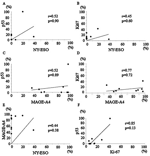
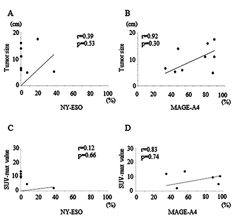
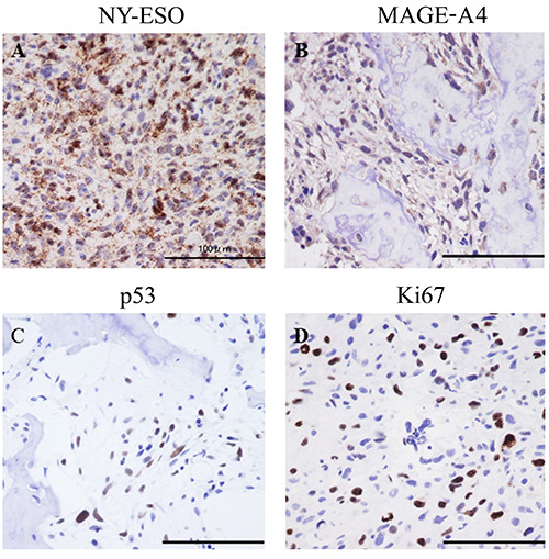
The cancer/testis antigens (CTAs), New York esophageal squamous cell carcinoma-1 (NY-ESO-1) and melanoma antigen gene (MAGE)-A4 are normally restricted to male germ cells but are aberrantly expressed in several cancers. Considering the limited information regarding their significance in osteosarcoma (OS), the purpose of this study was to determine the clinical significance of NY-ESO-1 and MAGE-A4 expression in OS. Nine patients with OS treated at Kindai University Hospital were included in the study. The median age was 27 years, and median follow-up period was 40 months. The specimens obtained at the time of biopsy were used to perform immunostaining for NY-ESO, MAGE-A4, p53, and Ki-67. The positive cell rates and positive case rates of NY-ESO, MAGE-A4, p53, and Ki-67 were calculated. The correlation between the positive cell rate of immunohistochemical markers was also calculated. The correlation between the positive cell rate of NY-ESO-1 or MAGE-A4 and tumor size or maximum standardized uptake (SUV-max) was also determined. The positive cell rates of NY-ESO-1 or MAGE-A4 in continuous disease-free (CDF) cases were also compared with those in alive with disease (AWD) or dead of disease (DOD) cases. The average positive cell rates of NY-ESO, MAGEA4, p53, and Ki-67 were 71.7%, 85.1%, 16.2%, and 14.7%, and their positive case rates were 33.3%, 100%, 44.4%, and 100%, respectively. The positivity rates of NY-ESO-1 and p53 were strongly correlated, whereas those of NY-ESO-1 and Ki-67 were moderately correlated. The MAGE-A4 and p53 positivity rates and the MAGE-A4 and Ki-67 positive cell rates were both strongly correlated. The NY-ESO-1 and MAGE-A4 positivity rates were moderately correlated. The positive correlation between the NY-ESO-1 positive cell rate and tumor size was medium, and that between the MAGE-A4 positivity rate and SUV-max was very strong. There was no significant difference in the positive cell rates of NY-ESO-1 or MAGE-A4 between CDF cases and AWD or DOD cases. Overall, our results suggest that NY-ESO-1 and MAGE-A4 may be involved in the aggressiveness of OS.
期刊介绍:
The Journal publishes original papers concerning investigations by histochemical and immunohistochemical methods, and performed with the aid of light, super-resolution and electron microscopy, cytometry and imaging techniques. Coverage extends to:
functional cell and tissue biology in animals and plants;
cell differentiation and death;
cell-cell interaction and molecular trafficking;
biology of cell development and senescence;
nerve and muscle cell biology;
cellular basis of diseases.
The histochemical approach is nowadays essentially aimed at locating molecules in the very place where they exert their biological roles, and at describing dynamically specific chemical activities in living cells. Basic research on cell functional organization is essential for understanding the mechanisms underlying major biological processes such as differentiation, the control of tissue homeostasis, and the regulation of normal and tumor cell growth. Even more than in the past, the European Journal of Histochemistry, as a journal of functional cytology, represents the venue where cell scientists may present and discuss their original results, technical improvements and theories.
 求助内容:
求助内容: 应助结果提醒方式:


