Comparative Evaluation of Clinical Efficacy of Leukocyte-Rich Platelet-Rich Fibrin with Advanced Platelet-Rich Fibrin in Management of Gingival Recession Defects: A Randomized Controlled Trial.
{"title":"Comparative Evaluation of Clinical Efficacy of Leukocyte-Rich Platelet-Rich Fibrin with Advanced Platelet-Rich Fibrin in Management of Gingival Recession Defects: A Randomized Controlled Trial.","authors":"Anupama Tadepalli, Swapna Chekurthi, Swarupa Kavassery Balasubramanian, Harinath Parthasarathy, Deepa Ponnaiyan","doi":"10.1159/000525560","DOIUrl":null,"url":null,"abstract":"<p><strong>Background: </strong>The aim of this research was to determine and compare the clinical efficacy of leukocyte platelet-rich fibrin (L-PRF) and advanced platelet-rich fibrin (A-PRF) in combination with coronally advanced flap (CAF) in the treatment of gingival recession defects.</p><p><strong>Methods: </strong>Systemically healthy subjects presenting with 30 Miller's class I or II gingival recession defects in maxillary anteriors and premolars, were treated with either CAF + L-PRF or CAF + A-PRF. Clinical parameters such as recession height (RH), width, probing pocket depth, clinical attachment level (CAL), keratinized tissue height (KTH), and width of attached gingiva (WAG) were measured at baseline, 3, and 6 months. Gingival biotype was evaluated at baseline and 6 months post-surgery. Mean root coverage percentage (MRC%) was evaluated at 3 and 6 months.</p><p><strong>Results: </strong>Statistically significant reduction in mean RH was observed from baseline (2.53 ± 0.74 mm, 2.63 ± 0.82 mm) to 6 months (0.87 ± 0.83 mm, 0.53 ± 0.91 mm) in CAF + L-PRF and CAF + A-PRF groups, respectively. The MRC% achieved at 6 months was 67.20 ± 32.81 in the CAF + L-PRF group and 81.66 ± 28.21 in the CAF + A-PRF group. Statistically significant gain in CAL, WAG, and KTH was observed in both therapeutic groups (p < 0.05). Intergroup analysis revealed no statistically significant differences among study parameters between groups at any time point (p > 0.05).</p><p><strong>Conclusion: </strong>Based on the findings of this study, both L-PRF and A-PRF may be suggested as viable treatment options for the management of gingival recession in maxilla.</p>","PeriodicalId":520708,"journal":{"name":"Medical principles and practice : international journal of the Kuwait University, Health Science Centre","volume":" ","pages":"376-383"},"PeriodicalIF":2.2000,"publicationDate":"2022-01-01","publicationTypes":"Journal Article","fieldsOfStudy":null,"isOpenAccess":false,"openAccessPdf":"https://ftp.ncbi.nlm.nih.gov/pub/pmc/oa_pdf/6b/35/mpp-0031-0376.PMC9485915.pdf","citationCount":"1","resultStr":null,"platform":"Semanticscholar","paperid":null,"PeriodicalName":"Medical principles and practice : international journal of the Kuwait University, Health Science Centre","FirstCategoryId":"3","ListUrlMain":"https://doi.org/10.1159/000525560","RegionNum":0,"RegionCategory":null,"ArticlePicture":[],"TitleCN":null,"AbstractTextCN":null,"PMCID":null,"EPubDate":"2022/6/21 0:00:00","PubModel":"Epub","JCR":"","JCRName":"","Score":null,"Total":0}
引用次数: 1
Abstract
Background: The aim of this research was to determine and compare the clinical efficacy of leukocyte platelet-rich fibrin (L-PRF) and advanced platelet-rich fibrin (A-PRF) in combination with coronally advanced flap (CAF) in the treatment of gingival recession defects.
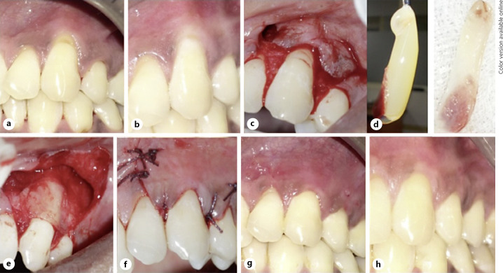
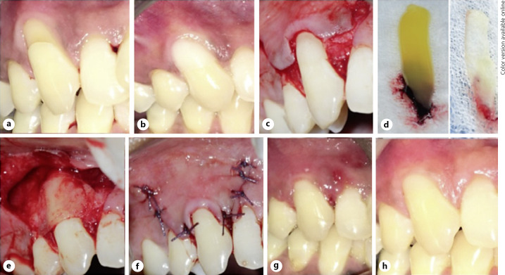
Methods: Systemically healthy subjects presenting with 30 Miller's class I or II gingival recession defects in maxillary anteriors and premolars, were treated with either CAF + L-PRF or CAF + A-PRF. Clinical parameters such as recession height (RH), width, probing pocket depth, clinical attachment level (CAL), keratinized tissue height (KTH), and width of attached gingiva (WAG) were measured at baseline, 3, and 6 months. Gingival biotype was evaluated at baseline and 6 months post-surgery. Mean root coverage percentage (MRC%) was evaluated at 3 and 6 months.
Results: Statistically significant reduction in mean RH was observed from baseline (2.53 ± 0.74 mm, 2.63 ± 0.82 mm) to 6 months (0.87 ± 0.83 mm, 0.53 ± 0.91 mm) in CAF + L-PRF and CAF + A-PRF groups, respectively. The MRC% achieved at 6 months was 67.20 ± 32.81 in the CAF + L-PRF group and 81.66 ± 28.21 in the CAF + A-PRF group. Statistically significant gain in CAL, WAG, and KTH was observed in both therapeutic groups (p < 0.05). Intergroup analysis revealed no statistically significant differences among study parameters between groups at any time point (p > 0.05).
Conclusion: Based on the findings of this study, both L-PRF and A-PRF may be suggested as viable treatment options for the management of gingival recession in maxilla.

 求助内容:
求助内容: 应助结果提醒方式:


