{"title":"Multimodal Imaging of Subfoveal Pachydrusen Containing a Blood Flow Signal.","authors":"Naoko Ishiguro, Takaaki Hayashi, Yoshiko Yamawaki, Kei Mizobuchi, Tsutomu Yasukawa, Shigeru Honda, Tadashi Nakano","doi":"10.1155/2022/5680913","DOIUrl":null,"url":null,"abstract":"<p><p>Individuals with pachydrusen, larger than 125 <i>μ</i>m, have a significantly thicker choroid than do those with soft drusen or reticular pseudodrusen. Little is known about cases of abnormal blood flow within pachydrusen. The purpose of this report was to demonstrate a blood flow signal within pachydrusen using optical coherence tomography (OCT) angiography. A 76-year-old Japanese woman presented with innumerable drusen/pachydrusen in both posterior poles. Her visual acuity was good. OCT showed subfoveal pachydrusen in the left eye, but no exudative changes. The subfoveal choroidal thickness was increased to 274 <i>μ</i>m in the left eye. OCT angiography revealed a blood flow signal within the pachydrusen. However, fluorescein and indocyanine green angiographies indicated no abnormal hyperfluorescent lesion in the macula of the left eye. During the 13-month follow-up, the blood flow signal in OCT angiography did not change in diameter, and no exudative change was observed. The blood flow signal may have properties of capillary blood vessels derived from the choriocapillaris, rather than angiogenic vessels from choroidal neovascularization or polypoidal choroidal vasculopathy/aneurysmal type 1 neovascularization.</p>","PeriodicalId":9603,"journal":{"name":"Case Reports in Ophthalmological Medicine","volume":" ","pages":"5680913"},"PeriodicalIF":0.4000,"publicationDate":"2022-06-08","publicationTypes":"Journal Article","fieldsOfStudy":null,"isOpenAccess":false,"openAccessPdf":"https://www.ncbi.nlm.nih.gov/pmc/articles/PMC9200588/pdf/","citationCount":"0","resultStr":null,"platform":"Semanticscholar","paperid":null,"PeriodicalName":"Case Reports in Ophthalmological Medicine","FirstCategoryId":"1085","ListUrlMain":"https://doi.org/10.1155/2022/5680913","RegionNum":0,"RegionCategory":null,"ArticlePicture":[],"TitleCN":null,"AbstractTextCN":null,"PMCID":null,"EPubDate":"2022/1/1 0:00:00","PubModel":"eCollection","JCR":"Q4","JCRName":"OPHTHALMOLOGY","Score":null,"Total":0}
引用次数: 0
Abstract
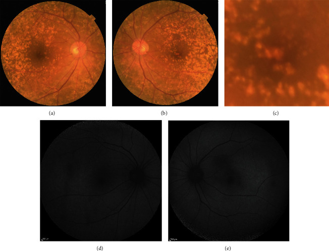
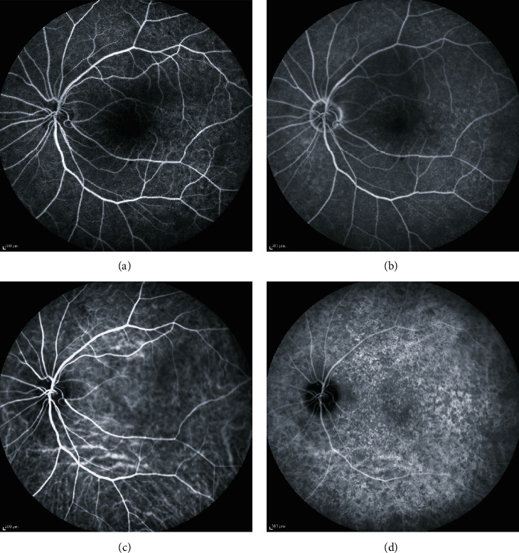
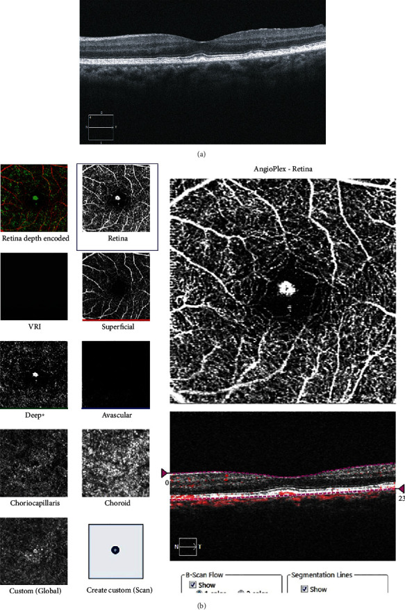
Individuals with pachydrusen, larger than 125 μm, have a significantly thicker choroid than do those with soft drusen or reticular pseudodrusen. Little is known about cases of abnormal blood flow within pachydrusen. The purpose of this report was to demonstrate a blood flow signal within pachydrusen using optical coherence tomography (OCT) angiography. A 76-year-old Japanese woman presented with innumerable drusen/pachydrusen in both posterior poles. Her visual acuity was good. OCT showed subfoveal pachydrusen in the left eye, but no exudative changes. The subfoveal choroidal thickness was increased to 274 μm in the left eye. OCT angiography revealed a blood flow signal within the pachydrusen. However, fluorescein and indocyanine green angiographies indicated no abnormal hyperfluorescent lesion in the macula of the left eye. During the 13-month follow-up, the blood flow signal in OCT angiography did not change in diameter, and no exudative change was observed. The blood flow signal may have properties of capillary blood vessels derived from the choriocapillaris, rather than angiogenic vessels from choroidal neovascularization or polypoidal choroidal vasculopathy/aneurysmal type 1 neovascularization.

 求助内容:
求助内容: 应助结果提醒方式:


