{"title":"Sputum characteristics of patients with severe COVID-19: report of two cases with immunocytochemical detection of SARS-CoV-2 spike protein.","authors":"Daichi Fujimoto, Minako Fukuya, Sachie Terao, Isao Irei, Takashi Akiyama, Anna Watanabe, Yuri Yasuda, Daisuke Yoshioka, Kazuhide Takada, Satoshi Hayakawa, Takuya Moriya","doi":"10.1007/s00795-022-00326-9","DOIUrl":null,"url":null,"abstract":"<p><p>Patients with SARS-CoV-2 infection and with severe COVID-19 often have multiple coinfections, and their treatment is challenging. Here, we performed cytology analysis on sputum samples from two patients with severe COVID-19. The specimens were prepared using the rubbing method and stained with Papanicolaou stain. In both cases, several cells with frosted nuclei were observed, and the cytological findings per 100 cells were evaluated. The infected cells were mononuclear to multinuclear, showing chromatin aggregation at the nuclear margins, intranuclear inclusion bodies, eosinophilic cytoplasmic inclusion bodies, and mutual pressure exclusion of the nuclei. Immunocytochemical staining revealed that the cells were positive for AE1/AE3 and negative for CD68 expression, indicating their epithelial origin. Furthermore, infected cells with frosted nuclei were positive for surfactant protein A (SP-A) in Case 2, suggesting infection of type II alveolar pneumocytes or Clara cells. Moreover, in Case 2, the infected cells were positive for herpes simplex virus (HSV) I + II and SARS-CoV-2 spike protein, confirming double infection in these cells. In conclusion, sputum cytology is an important tool for determining the diversity of viral infection, and additional immunocytochemistry can be used for definitive diagnosis.</p>","PeriodicalId":1,"journal":{"name":"Accounts of Chemical Research","volume":null,"pages":null},"PeriodicalIF":16.4000,"publicationDate":"2022-12-01","publicationTypes":"Journal Article","fieldsOfStudy":null,"isOpenAccess":false,"openAccessPdf":"https://www.ncbi.nlm.nih.gov/pmc/articles/PMC9206128/pdf/","citationCount":"2","resultStr":null,"platform":"Semanticscholar","paperid":null,"PeriodicalName":"Accounts of Chemical Research","FirstCategoryId":"3","ListUrlMain":"https://doi.org/10.1007/s00795-022-00326-9","RegionNum":1,"RegionCategory":"化学","ArticlePicture":[],"TitleCN":null,"AbstractTextCN":null,"PMCID":null,"EPubDate":"2022/6/18 0:00:00","PubModel":"Epub","JCR":"Q1","JCRName":"CHEMISTRY, MULTIDISCIPLINARY","Score":null,"Total":0}
引用次数: 2
Abstract
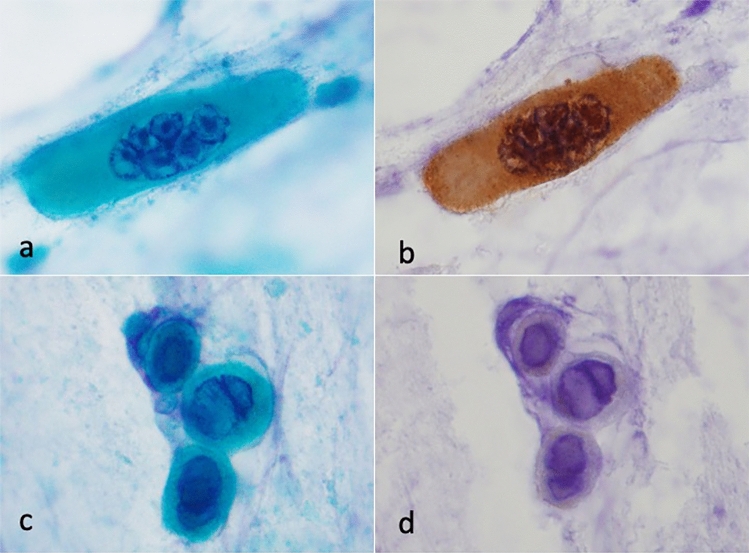
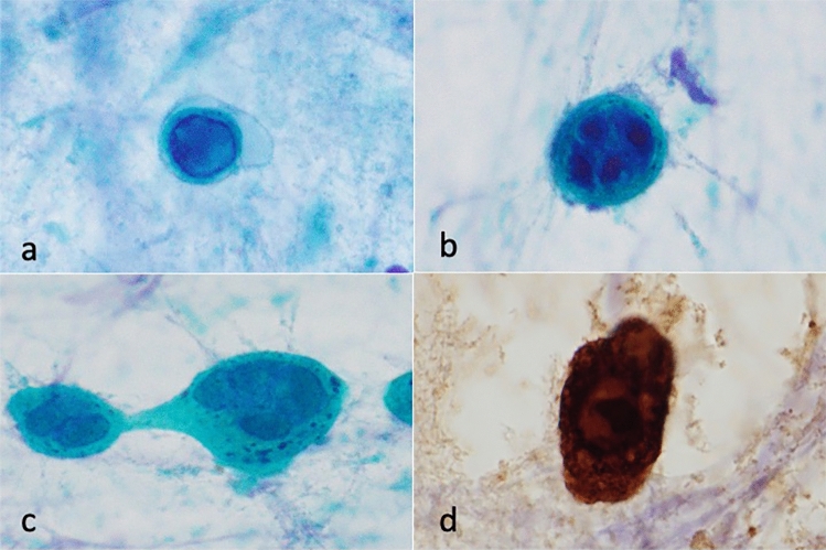
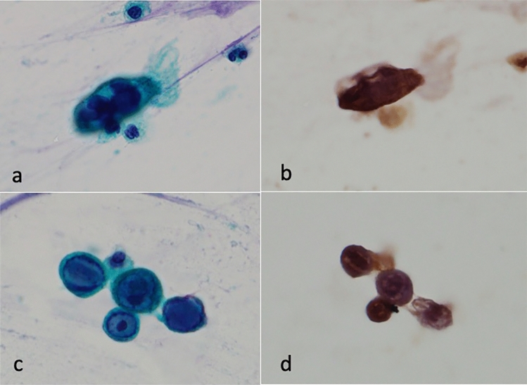
Patients with SARS-CoV-2 infection and with severe COVID-19 often have multiple coinfections, and their treatment is challenging. Here, we performed cytology analysis on sputum samples from two patients with severe COVID-19. The specimens were prepared using the rubbing method and stained with Papanicolaou stain. In both cases, several cells with frosted nuclei were observed, and the cytological findings per 100 cells were evaluated. The infected cells were mononuclear to multinuclear, showing chromatin aggregation at the nuclear margins, intranuclear inclusion bodies, eosinophilic cytoplasmic inclusion bodies, and mutual pressure exclusion of the nuclei. Immunocytochemical staining revealed that the cells were positive for AE1/AE3 and negative for CD68 expression, indicating their epithelial origin. Furthermore, infected cells with frosted nuclei were positive for surfactant protein A (SP-A) in Case 2, suggesting infection of type II alveolar pneumocytes or Clara cells. Moreover, in Case 2, the infected cells were positive for herpes simplex virus (HSV) I + II and SARS-CoV-2 spike protein, confirming double infection in these cells. In conclusion, sputum cytology is an important tool for determining the diversity of viral infection, and additional immunocytochemistry can be used for definitive diagnosis.
SARS-CoV-2感染患者和严重的COVID-19患者通常有多次合并感染,其治疗具有挑战性。在这里,我们对两名重症COVID-19患者的痰样本进行了细胞学分析。采用摩擦法制备标本,并用Papanicolaou染色。在这两种情况下,观察到几个细胞核结霜的细胞,并评估每100个细胞的细胞学结果。感染细胞为单核到多核,核边缘染色质聚集,核内包涵体,嗜酸性细胞质包涵体,细胞核相互压力排斥。免疫细胞化学染色显示细胞AE1/AE3表达阳性,CD68表达阴性,提示其上皮来源。此外,在病例2中,感染的细胞核呈霜状的细胞表面活性剂蛋白A (SP-A)阳性,提示感染了II型肺泡肺细胞或Clara细胞。此外,在病例2中,感染细胞对单纯疱疹病毒(HSV) I + II和SARS-CoV-2刺突蛋白呈阳性,证实这些细胞存在双重感染。总之,痰细胞学检查是确定病毒感染多样性的重要工具,另外的免疫细胞化学检查可用于明确诊断。
期刊介绍:
Accounts of Chemical Research presents short, concise and critical articles offering easy-to-read overviews of basic research and applications in all areas of chemistry and biochemistry. These short reviews focus on research from the author’s own laboratory and are designed to teach the reader about a research project. In addition, Accounts of Chemical Research publishes commentaries that give an informed opinion on a current research problem. Special Issues online are devoted to a single topic of unusual activity and significance.
Accounts of Chemical Research replaces the traditional article abstract with an article "Conspectus." These entries synopsize the research affording the reader a closer look at the content and significance of an article. Through this provision of a more detailed description of the article contents, the Conspectus enhances the article's discoverability by search engines and the exposure for the research.

 求助内容:
求助内容: 应助结果提醒方式:


