{"title":"Oligogenic cardiomyopathy.","authors":"Ali J Marian","doi":"10.20517/jca.2021.27","DOIUrl":null,"url":null,"abstract":"Dr. McKenna and colleagues interpret the histology figure to diagnose cor adiposum in family member III-4 who died suddenly and was found to have extensive fibro-adiposis of the right ventricle[1]. The authors apparently made their diagnosis indicating that fibrosis is necessary for the diagnosis of arrhythmogenic right ventricular cardiomyopathy (ARVC). We had described the histological data as “fibro-fatty infiltration of the right ventricle, encompassing 50% to 80% of the right ventricular wall thickness”[1]. However, we had not included specific staining for myocardial fibrosis. We provide Masson trichrome-stained myocardial sections, which show unequivocal evidence of myocardial fibrosis along with the excess adipocytes [Figure 1]. We also note that individual III-4 had pathogenic and likely pathogenic variants (PVs/LPVs) in the PKP2 and DSP genes, which are well-established causes of ARVC. Furthermore, cor adiposum does not exclusively and extensively involve the right ventricle, as observed in individual III-4, without involving the left ventricle. Thus, the data firmly refutes the diagnosis of cor adiposum and confirms the diagnosis of the classic ARVC.","PeriodicalId":75051,"journal":{"name":"The journal of cardiovascular aging","volume":" ","pages":""},"PeriodicalIF":0.0000,"publicationDate":"2022-01-01","publicationTypes":"Journal Article","fieldsOfStudy":null,"isOpenAccess":false,"openAccessPdf":"https://www.ncbi.nlm.nih.gov/pmc/articles/PMC8623865/pdf/","citationCount":"1","resultStr":null,"platform":"Semanticscholar","paperid":null,"PeriodicalName":"The journal of cardiovascular aging","FirstCategoryId":"1085","ListUrlMain":"https://doi.org/10.20517/jca.2021.27","RegionNum":0,"RegionCategory":null,"ArticlePicture":[],"TitleCN":null,"AbstractTextCN":null,"PMCID":null,"EPubDate":"","PubModel":"","JCR":"","JCRName":"","Score":null,"Total":0}
引用次数: 1
Abstract
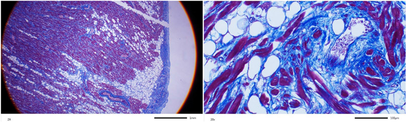
Dr. McKenna and colleagues interpret the histology figure to diagnose cor adiposum in family member III-4 who died suddenly and was found to have extensive fibro-adiposis of the right ventricle[1]. The authors apparently made their diagnosis indicating that fibrosis is necessary for the diagnosis of arrhythmogenic right ventricular cardiomyopathy (ARVC). We had described the histological data as “fibro-fatty infiltration of the right ventricle, encompassing 50% to 80% of the right ventricular wall thickness”[1]. However, we had not included specific staining for myocardial fibrosis. We provide Masson trichrome-stained myocardial sections, which show unequivocal evidence of myocardial fibrosis along with the excess adipocytes [Figure 1]. We also note that individual III-4 had pathogenic and likely pathogenic variants (PVs/LPVs) in the PKP2 and DSP genes, which are well-established causes of ARVC. Furthermore, cor adiposum does not exclusively and extensively involve the right ventricle, as observed in individual III-4, without involving the left ventricle. Thus, the data firmly refutes the diagnosis of cor adiposum and confirms the diagnosis of the classic ARVC.

 求助内容:
求助内容: 应助结果提醒方式:


