Glaucomatous Type Cupping Caused by Internal Carotid Artery Compression: a Case Report.
IF 2.8
Q2 CLINICAL NEUROLOGY
Journal of Central Nervous System Disease
Pub Date : 2022-02-14
eCollection Date: 2022-01-01
DOI:10.1177/11795735221081588
引用次数: 0
Abstract
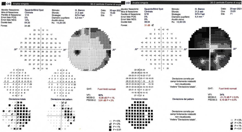
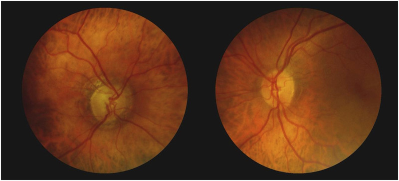
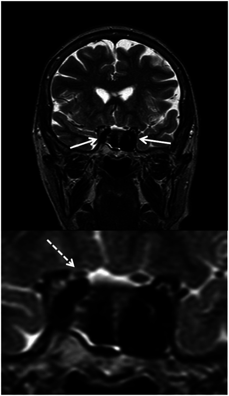
A 71-year-old woman with a diagnosis of normal tension glaucoma (NTG) presented with complains of progressive visual loss in the right eye. Examination revealed features consistent with compressive optic neuropathy. Although brain magnetic resonance imaging (MRI) was initially interpreted as normal, re-evaluation disclosed a compression on the right optic nerve from the right internal carotid artery. We highlight the clinical differential diagnosis between NTG and compressive optic neuropathy. This case is a reminder that a compressive optic neuropathy may be caused by anatomical variation of normal intracranial structures.


颈内动脉压迫致青光眼型拔罐1例。
一位71岁女性,诊断为正常紧张性青光眼(NTG),主诉右眼进行性视力丧失。检查显示与压迫性视神经病变相符。虽然脑磁共振成像(MRI)最初被解释为正常,但重新评估显示右侧颈内动脉压迫右侧视神经。我们强调NTG和压迫性视神经病变的临床鉴别诊断。这个病例提醒我们,压缩性视神经病变可能是由正常颅内结构的解剖变异引起的。
本文章由计算机程序翻译,如有差异,请以英文原文为准。
求助全文
约1分钟内获得全文
求助全文
来源期刊

Journal of Central Nervous System Disease
CLINICAL NEUROLOGY-
CiteScore
6.90
自引率
0.00%
发文量
39
审稿时长
8 weeks
 求助内容:
求助内容: 应助结果提醒方式:


