Predictors of worsening TR severity after right ventricular lead placement: any added value by post-procedural fluoroscopy versus three -dimensional echocardiography?
Hoorak Poorzand, Mohammad Tayyebi, Sara Hosseini, Alireza Heidari Bakavoli, Faeze Keihanian, Lida Jarahi, Ali Hamadanchi
{"title":"Predictors of worsening TR severity after right ventricular lead placement: any added value by post-procedural fluoroscopy versus three -dimensional echocardiography?","authors":"Hoorak Poorzand, Mohammad Tayyebi, Sara Hosseini, Alireza Heidari Bakavoli, Faeze Keihanian, Lida Jarahi, Ali Hamadanchi","doi":"10.1186/s12947-021-00267-w","DOIUrl":null,"url":null,"abstract":"<p><strong>Background: </strong>The effect of right ventricular (RV) leads on tricuspid valve has been already raised concerns, especially in terms of prognostic implication. For such assessment, three-dimensional transthoracic echocardiography (3D-TTE) has been used previously but there was no data on the use of post-procedural fluoroscopy in the literature.</p><p><strong>Methods: </strong>We prospectively enrolled 59 patients who underwent clinically indicated placement of pacemaker or implantable cardioverter defibrillator (ICD). Vena contracta (VC) and tricuspid regurgitation (TR) severity were measured using two-dimensional transthoracic echocardiography (2D-TTE) at baseline. Follow up 3D-TTE was performed 6 months after device implantation to assess TR severity and RV lead location.</p><p><strong>Results: </strong>Lead placement position in TV was defined in 51 cases.TR VC was increased after the lead placement, compared to the baseline study (VC: 3.86 ± 2.32 vs 3.18 ± 2.39; p = 0.005), with one grade worsening in TR in 25.4% of cases. The mean changes in VC levels were 1.14 ± 0.67 mm. Among all investigated parameters, VC changes were predicted based on lead placement position only in 3D-TTE (p < 0.001) while the other variables including fluoroscopy parameters were not informative.</p><p><strong>Conclusion: </strong>The RV Lead location examined by 3D-TTE seems to be a valuable parameter to predict the changes in the severity of the tricuspid regurgitation. Fluoroscopy findings did not improve the predictive performance, at least in short term follow up.</p>","PeriodicalId":9613,"journal":{"name":"Cardiovascular Ultrasound","volume":"19 1","pages":"37"},"PeriodicalIF":1.6000,"publicationDate":"2021-11-21","publicationTypes":"Journal Article","fieldsOfStudy":null,"isOpenAccess":false,"openAccessPdf":"https://www.ncbi.nlm.nih.gov/pmc/articles/PMC8606093/pdf/","citationCount":"3","resultStr":null,"platform":"Semanticscholar","paperid":null,"PeriodicalName":"Cardiovascular Ultrasound","FirstCategoryId":"3","ListUrlMain":"https://doi.org/10.1186/s12947-021-00267-w","RegionNum":3,"RegionCategory":"医学","ArticlePicture":[],"TitleCN":null,"AbstractTextCN":null,"PMCID":null,"EPubDate":"","PubModel":"","JCR":"Q3","JCRName":"CARDIAC & CARDIOVASCULAR SYSTEMS","Score":null,"Total":0}
引用次数: 3
Abstract
Background: The effect of right ventricular (RV) leads on tricuspid valve has been already raised concerns, especially in terms of prognostic implication. For such assessment, three-dimensional transthoracic echocardiography (3D-TTE) has been used previously but there was no data on the use of post-procedural fluoroscopy in the literature.
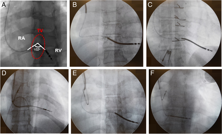
Methods: We prospectively enrolled 59 patients who underwent clinically indicated placement of pacemaker or implantable cardioverter defibrillator (ICD). Vena contracta (VC) and tricuspid regurgitation (TR) severity were measured using two-dimensional transthoracic echocardiography (2D-TTE) at baseline. Follow up 3D-TTE was performed 6 months after device implantation to assess TR severity and RV lead location.
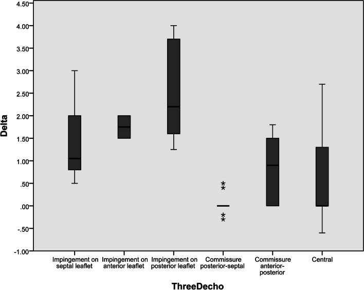
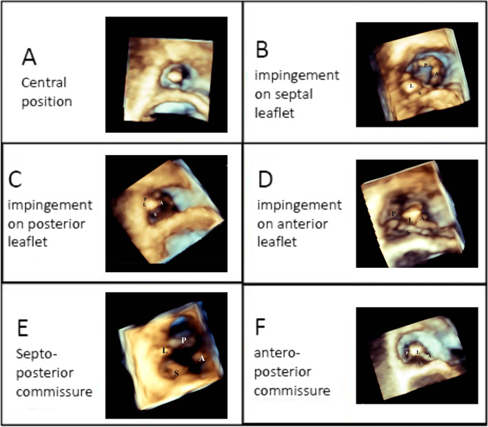
Results: Lead placement position in TV was defined in 51 cases.TR VC was increased after the lead placement, compared to the baseline study (VC: 3.86 ± 2.32 vs 3.18 ± 2.39; p = 0.005), with one grade worsening in TR in 25.4% of cases. The mean changes in VC levels were 1.14 ± 0.67 mm. Among all investigated parameters, VC changes were predicted based on lead placement position only in 3D-TTE (p < 0.001) while the other variables including fluoroscopy parameters were not informative.
Conclusion: The RV Lead location examined by 3D-TTE seems to be a valuable parameter to predict the changes in the severity of the tricuspid regurgitation. Fluoroscopy findings did not improve the predictive performance, at least in short term follow up.
期刊介绍:
Cardiovascular Ultrasound is an online journal, publishing peer-reviewed: original research; authoritative reviews; case reports on challenging and/or unusual diagnostic aspects; and expert opinions on new techniques and technologies. We are particularly interested in articles that include relevant images or video files, which provide an additional dimension to published articles and enhance understanding.
As an open access journal, Cardiovascular Ultrasound ensures high visibility for authors in addition to providing an up-to-date and freely available resource for the community. The journal welcomes discussion, and provides a forum for publishing opinion and debate ranging from biology to engineering to clinical echocardiography, with both speed and versatility.
 求助内容:
求助内容: 应助结果提醒方式:


