{"title":"Thoracic CT radiomics analysis for predicting synchronous brain metastasis in patients with lung cancer.","authors":"Zhimin Ding, Yuancheng Wang, Cong Xia, Xiangpan Meng, Qian Yu, Shenghong Ju","doi":"10.5152/dir.2021.21677","DOIUrl":null,"url":null,"abstract":"<p><strong>Purpose: </strong>We aimed to assess the feasibility of radiomics analysis based on non-contrast-enhanced thoracic CT images in predicting synchronous brain metastasis (SBM) in lung cancer patients at initial diagnosis.</p><p><strong>Methods: </strong>This retrospective study enrolled 371 lung cancer patients (with SBM n=147, without SBM n=224) confirmed by histopathology. Patients were allocated to the training set (n=258) and testing set (n=113). The optimal radiomics features were selected by using the least absolute shrinkage and selection operator (LASSO) algorithm. The radiomics, clinicoradiologic, and combined models were developed to predict SBM using multivariable logistic regression. Then the discrimination ability of the models was assessed. Furthermore, the prediction performance of the abovementioned three models for oligometastatic (1-3 lesions) or multiple (>3 lesions) brain metastases in SBM, metachronous brain metastasis (MBM), and total (SBM and MBM) groups were investigated.</p><p><strong>Results: </strong>Six radiomics features and two clinicoradiologic characteristics were chosen for predicting SBM. Both the radiomics model (area under the receiver operating characteristic curve [AUC] = 0.870 and 0.824 in the training and testing sets, respectively) and the combined model (AUC = 0.912 and 0.859, respectively) presented better predictive ability for SBM than the clinicoradiologic model (AUC = 0.712 and 0.692, respectively). The decision curve analysis (DCA) demonstrated the clinical usefulness of the radiomics-based models. The radiomics model can also be used to predict oligometastatic or multiple brain metastases in SBM, MBM, and total groups (P = .045, P = .022, and P = .030, respectively).</p><p><strong>Conclusion: </strong>The radiomics model and the combined model we presented can be used as valuable imaging markers for predicting patients at high risk of SBM at the initial diagnosis of lung cancer. Furthermore, the radiomics model can also be utilized as an indicator for identifying oligometastatic or multiple brain metastases.</p>","PeriodicalId":50582,"journal":{"name":"Diagnostic and Interventional Radiology","volume":" ","pages":"39-49"},"PeriodicalIF":1.7000,"publicationDate":"2022-01-01","publicationTypes":"Journal Article","fieldsOfStudy":null,"isOpenAccess":false,"openAccessPdf":"https://www.ncbi.nlm.nih.gov/pmc/articles/PMC12278919/pdf/","citationCount":"0","resultStr":null,"platform":"Semanticscholar","paperid":null,"PeriodicalName":"Diagnostic and Interventional Radiology","FirstCategoryId":"3","ListUrlMain":"https://doi.org/10.5152/dir.2021.21677","RegionNum":4,"RegionCategory":"医学","ArticlePicture":[],"TitleCN":null,"AbstractTextCN":null,"PMCID":null,"EPubDate":"","PubModel":"","JCR":"Q2","JCRName":"Medicine","Score":null,"Total":0}
引用次数: 0
Abstract
Purpose: We aimed to assess the feasibility of radiomics analysis based on non-contrast-enhanced thoracic CT images in predicting synchronous brain metastasis (SBM) in lung cancer patients at initial diagnosis.
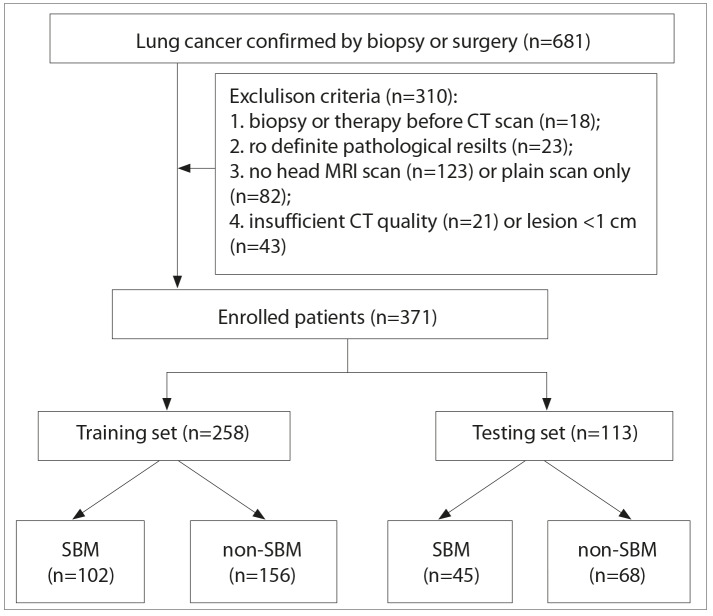
Methods: This retrospective study enrolled 371 lung cancer patients (with SBM n=147, without SBM n=224) confirmed by histopathology. Patients were allocated to the training set (n=258) and testing set (n=113). The optimal radiomics features were selected by using the least absolute shrinkage and selection operator (LASSO) algorithm. The radiomics, clinicoradiologic, and combined models were developed to predict SBM using multivariable logistic regression. Then the discrimination ability of the models was assessed. Furthermore, the prediction performance of the abovementioned three models for oligometastatic (1-3 lesions) or multiple (>3 lesions) brain metastases in SBM, metachronous brain metastasis (MBM), and total (SBM and MBM) groups were investigated.
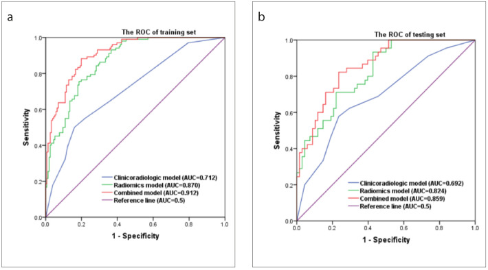
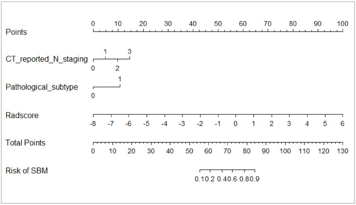
Results: Six radiomics features and two clinicoradiologic characteristics were chosen for predicting SBM. Both the radiomics model (area under the receiver operating characteristic curve [AUC] = 0.870 and 0.824 in the training and testing sets, respectively) and the combined model (AUC = 0.912 and 0.859, respectively) presented better predictive ability for SBM than the clinicoradiologic model (AUC = 0.712 and 0.692, respectively). The decision curve analysis (DCA) demonstrated the clinical usefulness of the radiomics-based models. The radiomics model can also be used to predict oligometastatic or multiple brain metastases in SBM, MBM, and total groups (P = .045, P = .022, and P = .030, respectively).
Conclusion: The radiomics model and the combined model we presented can be used as valuable imaging markers for predicting patients at high risk of SBM at the initial diagnosis of lung cancer. Furthermore, the radiomics model can also be utilized as an indicator for identifying oligometastatic or multiple brain metastases.
期刊介绍:
Diagnostic and Interventional Radiology (Diagn Interv Radiol) is the open access, online-only official publication of Turkish Society of Radiology. It is published bimonthly and the journal’s publication language is English.
The journal is a medium for original articles, reviews, pictorial essays, technical notes related to all fields of diagnostic and interventional radiology.

 求助内容:
求助内容: 应助结果提醒方式:


