Orwa Aboud, Ritu Shah, Elizabeth Vera, Eric Burton, Brett Theeler, Jing Wu, Lisa Boris, Martha Quezado, Jennifer Reyes, Kathleen Wall, Mark R Gilbert, Terri S Armstrong, Marta Penas-Prado
{"title":"Challenges of imaging interpretation to predict oligodendroglioma grade: a report from the Neuro-Oncology Branch.","authors":"Orwa Aboud, Ritu Shah, Elizabeth Vera, Eric Burton, Brett Theeler, Jing Wu, Lisa Boris, Martha Quezado, Jennifer Reyes, Kathleen Wall, Mark R Gilbert, Terri S Armstrong, Marta Penas-Prado","doi":"10.2217/cns-2021-0005","DOIUrl":null,"url":null,"abstract":"<p><p><b>Background:</b> To illustrate challenges of imaging interpretation in patients with oligodendroglioma seen at a referral center and evaluate interrater reliability. <b>Methods:</b> Two neuro-oncologists reviewed diagnostic preradiation MRIs of oligodendroglioma patients; interrater reliability was calculated with the kappa coefficient (k). A neuroradiologist measured presurgical apparent diffusion coefficient (ADC), if available. <b>Results:</b> Extensive enhancement was noted in four of 58 patients, k = 0.7; necrosis in seven of 58, k = 0.61; calcification in seven of 17, k = 1.0; diffusion restriction in two of 39 patients, k = 1.0 (all only in grade 3). ADC values with receiver operator characteristic analysis for area under the curve were 0.473, not significantly different from the null hypothesis (p = 0.14). <b>Conclusions:</b> Extensive enhancement, necrosis and calcification correlated with grade 3 oligodendroglioma in our sample. However, interrater variability is an important limitation when assessing radiographic features, supporting the need for standardization of imaging protocols and their interpretation.</p>","PeriodicalId":10469,"journal":{"name":"CNS Oncology","volume":" ","pages":"CNS83"},"PeriodicalIF":0.0000,"publicationDate":"2022-03-01","publicationTypes":"Journal Article","fieldsOfStudy":null,"isOpenAccess":false,"openAccessPdf":"https://ftp.ncbi.nlm.nih.gov/pub/pmc/oa_pdf/16/3e/cns-11-83.PMC8988255.pdf","citationCount":"4","resultStr":null,"platform":"Semanticscholar","paperid":null,"PeriodicalName":"CNS Oncology","FirstCategoryId":"1085","ListUrlMain":"https://doi.org/10.2217/cns-2021-0005","RegionNum":0,"RegionCategory":null,"ArticlePicture":[],"TitleCN":null,"AbstractTextCN":null,"PMCID":null,"EPubDate":"2022/2/10 0:00:00","PubModel":"Epub","JCR":"Q1","JCRName":"Medicine","Score":null,"Total":0}
引用次数: 4
Abstract
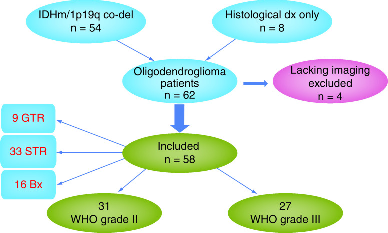
Background: To illustrate challenges of imaging interpretation in patients with oligodendroglioma seen at a referral center and evaluate interrater reliability. Methods: Two neuro-oncologists reviewed diagnostic preradiation MRIs of oligodendroglioma patients; interrater reliability was calculated with the kappa coefficient (k). A neuroradiologist measured presurgical apparent diffusion coefficient (ADC), if available. Results: Extensive enhancement was noted in four of 58 patients, k = 0.7; necrosis in seven of 58, k = 0.61; calcification in seven of 17, k = 1.0; diffusion restriction in two of 39 patients, k = 1.0 (all only in grade 3). ADC values with receiver operator characteristic analysis for area under the curve were 0.473, not significantly different from the null hypothesis (p = 0.14). Conclusions: Extensive enhancement, necrosis and calcification correlated with grade 3 oligodendroglioma in our sample. However, interrater variability is an important limitation when assessing radiographic features, supporting the need for standardization of imaging protocols and their interpretation.

 求助内容:
求助内容: 应助结果提醒方式:


