Comparison of Electrical Impedance Tomography and Ultrasonography for Determination of Solid and Cystic Lesion Resembling Breast Tumor Embedded in Chicken Phantom.
L Choridah, D Kurniadi, K Ain, M F Ulum, U Mukhaiyar, A D Garnadi, N H Setyawan
{"title":"Comparison of Electrical Impedance Tomography and Ultrasonography for Determination of Solid and Cystic Lesion Resembling Breast Tumor Embedded in Chicken Phantom.","authors":"L Choridah, D Kurniadi, K Ain, M F Ulum, U Mukhaiyar, A D Garnadi, N H Setyawan","doi":"10.2478/joeb-2021-0008","DOIUrl":null,"url":null,"abstract":"<p><p>Ultrasonography (US) and Electrical Impedance Tomography (EIT) can be used to detect breast cancer. Ultrasonography is based on non-ionizing radiations without adverse biological effects. A set of electrodes was placed around the torso and a small alternating current (AC) was injected via two of the electrodes into the object. This study aimed to acquire preliminary data to evaluate the EIT method for differentiation of artificial solid and cystic tumors in comparison to standard US. This study used a phantom made from chicken meat. In order to obtain the image of the solid tumor, an olive with carrot insertion was done, and the cystic tumor was created by filling a small balloon with water. GE Logic C5 ultrasound was performed with a 12 MHz linear transducer. For EIT measurement, 16 ECG electrodes and 32 ECG electrodes were placed. Data processing was done using the Graz consensus Reconstruction algorithm for EIT (GREIT) and Newton's One Step Error Reconstructor (NOSER) methods. The artificial solid tumor produced an ultrasound image of an oval, inhomogeneous lesions. The GREIT method with 16 electrodes of artificial solid tumor did not show a match between the reconstructed image and the original object containing 2 anomalies, but a match was found with 32 electrodes. In the NOSER method, both 16 and 32 electrodes showed a match. Ultrasound of the artificial cystic tumor showed an oval, circumscribed, anechoic with posterior enhancement. Both the GREIT and NOSER methods using the artificial cystic tumor showed a match between the reconstructed image and the original object containing two anomalies. EIT has a lower imaging resolution in comparison to ultrasonography, but is progressively maturing as a tool for monitoring and imaging. The solid and cystic anomalies on the phantoms were visualized by the GREIT and NOSER methods except for the solid anomaly with the GREIT 16 electrodes.</p>","PeriodicalId":38125,"journal":{"name":"Journal of Electrical Bioimpedance","volume":"12 1","pages":"63-68"},"PeriodicalIF":0.0000,"publicationDate":"2021-11-20","publicationTypes":"Journal Article","fieldsOfStudy":null,"isOpenAccess":false,"openAccessPdf":"https://www.ncbi.nlm.nih.gov/pmc/articles/PMC8667814/pdf/","citationCount":"3","resultStr":null,"platform":"Semanticscholar","paperid":null,"PeriodicalName":"Journal of Electrical Bioimpedance","FirstCategoryId":"1085","ListUrlMain":"https://doi.org/10.2478/joeb-2021-0008","RegionNum":0,"RegionCategory":null,"ArticlePicture":[],"TitleCN":null,"AbstractTextCN":null,"PMCID":null,"EPubDate":"2021/1/1 0:00:00","PubModel":"eCollection","JCR":"Q3","JCRName":"Biochemistry, Genetics and Molecular Biology","Score":null,"Total":0}
引用次数: 3
Abstract
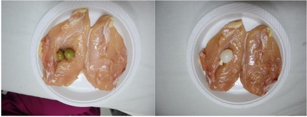
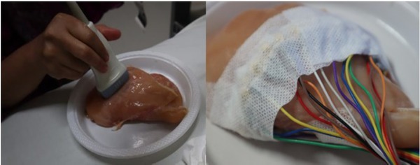
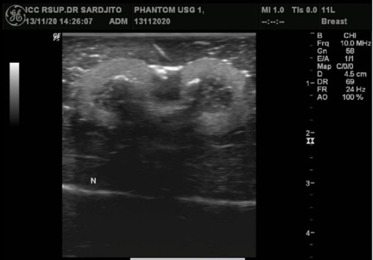
Ultrasonography (US) and Electrical Impedance Tomography (EIT) can be used to detect breast cancer. Ultrasonography is based on non-ionizing radiations without adverse biological effects. A set of electrodes was placed around the torso and a small alternating current (AC) was injected via two of the electrodes into the object. This study aimed to acquire preliminary data to evaluate the EIT method for differentiation of artificial solid and cystic tumors in comparison to standard US. This study used a phantom made from chicken meat. In order to obtain the image of the solid tumor, an olive with carrot insertion was done, and the cystic tumor was created by filling a small balloon with water. GE Logic C5 ultrasound was performed with a 12 MHz linear transducer. For EIT measurement, 16 ECG electrodes and 32 ECG electrodes were placed. Data processing was done using the Graz consensus Reconstruction algorithm for EIT (GREIT) and Newton's One Step Error Reconstructor (NOSER) methods. The artificial solid tumor produced an ultrasound image of an oval, inhomogeneous lesions. The GREIT method with 16 electrodes of artificial solid tumor did not show a match between the reconstructed image and the original object containing 2 anomalies, but a match was found with 32 electrodes. In the NOSER method, both 16 and 32 electrodes showed a match. Ultrasound of the artificial cystic tumor showed an oval, circumscribed, anechoic with posterior enhancement. Both the GREIT and NOSER methods using the artificial cystic tumor showed a match between the reconstructed image and the original object containing two anomalies. EIT has a lower imaging resolution in comparison to ultrasonography, but is progressively maturing as a tool for monitoring and imaging. The solid and cystic anomalies on the phantoms were visualized by the GREIT and NOSER methods except for the solid anomaly with the GREIT 16 electrodes.

 求助内容:
求助内容: 应助结果提醒方式:


