K A Bannikova, Yu Yu Bosykh, V G Gaitova, P G Sysolyatin, S P Sysolyatin
{"title":"Indications for the Use of Sialoendoscopy in Sialolithiasis.","authors":"K A Bannikova, Yu Yu Bosykh, V G Gaitova, P G Sysolyatin, S P Sysolyatin","doi":"10.17691/stm2020.12.3.05","DOIUrl":null,"url":null,"abstract":"<p><p><b>The aim of the study</b> is to determine indications for the use of sialoendoscopy in the diagnosis and treatment of sialolithiasis.</p><p><strong>Materials and methods: </strong>The study involved 115 patients with sialolithiasis, who underwent cone beam computed tomography, ultrasound diagnosis of the salivary glands, and sialoendoscopy, in addition to the standard general clinical examination.</p><p><strong>Results: </strong>Sialoendoscopy makes it possible to detect a stone, determine its shape, relative size, mobility, and assess the condition of the salivary ducts. It is impossible to obtain this information by other methods, though it is very important for treatment decision making. The design of the sialoscope and its special instruments make it possible to proceed with sialolith extraction immediately after detecting it.</p><p><strong>Conclusion: </strong>The absolute indication for the use of sialoendoscopy is mobile calculi less than 5 mm in diameter (L1 according to F. Marchal's LSD classification). In case of immobile sialoliths less than 4-8 mm in size, located in the main duct (L2), endoscopy should be used as a method supplementary to ductotomy. When sialoliths are located in the distal parts behind the areas of bending or stricture (L3a and L3b), the use of endoscopy is not indicated.</p>","PeriodicalId":51886,"journal":{"name":"Sovremennye Tehnologii v Medicine","volume":"12 3","pages":"41-45"},"PeriodicalIF":0.9000,"publicationDate":"2021-01-01","publicationTypes":"Journal Article","fieldsOfStudy":null,"isOpenAccess":false,"openAccessPdf":"https://www.ncbi.nlm.nih.gov/pmc/articles/PMC8596244/pdf/","citationCount":"1","resultStr":null,"platform":"Semanticscholar","paperid":null,"PeriodicalName":"Sovremennye Tehnologii v Medicine","FirstCategoryId":"1085","ListUrlMain":"https://doi.org/10.17691/stm2020.12.3.05","RegionNum":0,"RegionCategory":null,"ArticlePicture":[],"TitleCN":null,"AbstractTextCN":null,"PMCID":null,"EPubDate":"2020/6/28 0:00:00","PubModel":"Epub","JCR":"Q4","JCRName":"MEDICINE, RESEARCH & EXPERIMENTAL","Score":null,"Total":0}
引用次数: 1
Abstract
The aim of the study is to determine indications for the use of sialoendoscopy in the diagnosis and treatment of sialolithiasis.
Materials and methods: The study involved 115 patients with sialolithiasis, who underwent cone beam computed tomography, ultrasound diagnosis of the salivary glands, and sialoendoscopy, in addition to the standard general clinical examination.
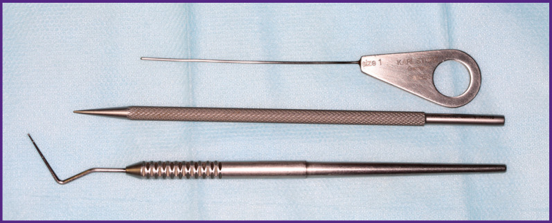
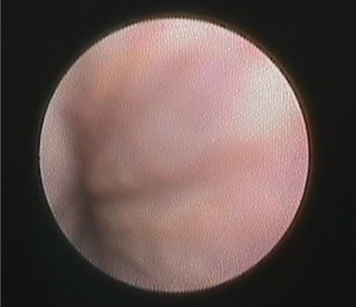
Results: Sialoendoscopy makes it possible to detect a stone, determine its shape, relative size, mobility, and assess the condition of the salivary ducts. It is impossible to obtain this information by other methods, though it is very important for treatment decision making. The design of the sialoscope and its special instruments make it possible to proceed with sialolith extraction immediately after detecting it.
Conclusion: The absolute indication for the use of sialoendoscopy is mobile calculi less than 5 mm in diameter (L1 according to F. Marchal's LSD classification). In case of immobile sialoliths less than 4-8 mm in size, located in the main duct (L2), endoscopy should be used as a method supplementary to ductotomy. When sialoliths are located in the distal parts behind the areas of bending or stricture (L3a and L3b), the use of endoscopy is not indicated.


 求助内容:
求助内容: 应助结果提醒方式:


