{"title":"Validation of the dosimetric and geometric accuracy of MR-only treatment planning solution for prostate cancer radiotherapy.","authors":"Michał Posiewnik, Tomasz Piotrowski","doi":"10.5114/wo.2021.112518","DOIUrl":null,"url":null,"abstract":"<p><strong>Introduction: </strong>The aim was to validate the dosimetric and geometric accuracy of radiotherapy treatment plans for prostate cancer based on magnetic resonance (MR) imaging only and a solution based on computed tomography (CT) supported by MR imaging.</p><p><strong>Material and methods: </strong>We used CT and MR images of ten prostate cancer patients implanted with three fiducial markers (FM) in the prostate gland. Rigid registration based on FM was performed to assess the fusion accuracy between MR and CT images. The differences between prostate contours (clinical target volume - CTV) on CT (CTV<sub>CT</sub>) and MR (CTV<sub>MR</sub>) images were scored using the Dice similarity coefficient and directly comparing the outlined volumes. The volumetric modulated arc therapy plans were designed and optimised on synthetic CT (sCT) to obtain the dose distribution for the MR-only solution. In the next step, the sCT images were replaced by conventional CT images and the plans were recalculated. The doses obtained on sCT and CT were compared by direct dose subtraction and the gamma method.</p><p><strong>Results: </strong>The averaged fiducial registration error was equal to 0.5 mm. All CTV<sub>CT</sub> volumes were significantly bigger than corresponding CTV delineated on MR images (<i>p</i> = 0.005). The direct dose comparison shows that for 97.1% of patients' bodies, the differences were smaller than 0.1%. The average gamma passing rates were higher than 0.970.</p><p><strong>Conclusions: </strong>MR imaging allows for a more precise delineation of the prostate compared to CT imaging. The workflow of plan preparation based on MR and CT is burdened with an FM registration error that is eliminated by an MR-only solution with no compromise on dose distribution.</p>","PeriodicalId":520599,"journal":{"name":"Contemporary oncology (Poznan, Poland)","volume":" ","pages":"249-253"},"PeriodicalIF":1.3000,"publicationDate":"2021-01-01","publicationTypes":"Journal Article","fieldsOfStudy":null,"isOpenAccess":false,"openAccessPdf":"https://ftp.ncbi.nlm.nih.gov/pub/pmc/oa_pdf/3f/60/WO-25-46150.PMC8768054.pdf","citationCount":"1","resultStr":null,"platform":"Semanticscholar","paperid":null,"PeriodicalName":"Contemporary oncology (Poznan, Poland)","FirstCategoryId":"1085","ListUrlMain":"https://doi.org/10.5114/wo.2021.112518","RegionNum":0,"RegionCategory":null,"ArticlePicture":[],"TitleCN":null,"AbstractTextCN":null,"PMCID":null,"EPubDate":"2022/1/5 0:00:00","PubModel":"Epub","JCR":"","JCRName":"","Score":null,"Total":0}
引用次数: 1
Abstract
Introduction: The aim was to validate the dosimetric and geometric accuracy of radiotherapy treatment plans for prostate cancer based on magnetic resonance (MR) imaging only and a solution based on computed tomography (CT) supported by MR imaging.
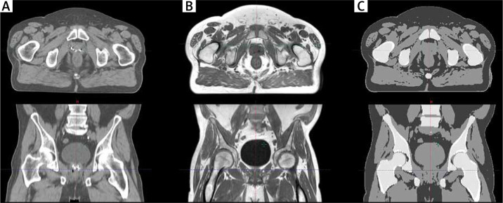
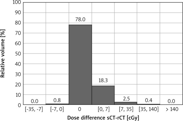
Material and methods: We used CT and MR images of ten prostate cancer patients implanted with three fiducial markers (FM) in the prostate gland. Rigid registration based on FM was performed to assess the fusion accuracy between MR and CT images. The differences between prostate contours (clinical target volume - CTV) on CT (CTVCT) and MR (CTVMR) images were scored using the Dice similarity coefficient and directly comparing the outlined volumes. The volumetric modulated arc therapy plans were designed and optimised on synthetic CT (sCT) to obtain the dose distribution for the MR-only solution. In the next step, the sCT images were replaced by conventional CT images and the plans were recalculated. The doses obtained on sCT and CT were compared by direct dose subtraction and the gamma method.
Results: The averaged fiducial registration error was equal to 0.5 mm. All CTVCT volumes were significantly bigger than corresponding CTV delineated on MR images (p = 0.005). The direct dose comparison shows that for 97.1% of patients' bodies, the differences were smaller than 0.1%. The average gamma passing rates were higher than 0.970.
Conclusions: MR imaging allows for a more precise delineation of the prostate compared to CT imaging. The workflow of plan preparation based on MR and CT is burdened with an FM registration error that is eliminated by an MR-only solution with no compromise on dose distribution.

 求助内容:
求助内容: 应助结果提醒方式:


