Immunoexpression of PD-L1, CD4+ and CD8+ cell infiltrates and tumor-infiltrating lymphocytes (TILs) in the microenvironment of actinic cheilitis and lower lip squamous cell carcinoma.
Vinícius Gonçalves de Souza, Damilys Joelly Souza Santos, Ana Gabriela Silva, Rosy Iara Maciel de Azambuja Ribeiro, Adriano Mota Loyola, Sérgio Vitorino Cardoso, Carla Silva Siqueira Miranda, Ludimila Paula Vaz Cardoso
{"title":"Immunoexpression of PD-L1, CD4+ and CD8+ cell infiltrates and tumor-infiltrating lymphocytes (TILs) in the microenvironment of actinic cheilitis and lower lip squamous cell carcinoma.","authors":"Vinícius Gonçalves de Souza, Damilys Joelly Souza Santos, Ana Gabriela Silva, Rosy Iara Maciel de Azambuja Ribeiro, Adriano Mota Loyola, Sérgio Vitorino Cardoso, Carla Silva Siqueira Miranda, Ludimila Paula Vaz Cardoso","doi":"10.1590/1678-7757-2021-0344","DOIUrl":null,"url":null,"abstract":"<p><strong>Objective: </strong>Lower lip squamous cell carcinomas (LLSCC) could be associated with a previous history of potentially malignant oral diseases (PMOD), especially actinic cheilitis (AC), with high sun exposure being a well-described risk factor. Immune evasion mechanisms, such as the PD-1/PD-L1 (programmed cell death protein 1/programmed death-ligand 1) pathway has been gaining prominence since immunotherapy with immune checkpoint inhibitors showed a positive effect on the survival of patients with different types of neoplasms. Concomitant with the characterization of the tumor microenvironment, the expression of either or both PD-1 and PD-L1 molecules may estimate mutual relations of progression or regression of the carcinoma and prognostic values of the patient.Considering the importance of tumor microenvironment characterization, this study aims to determine the immunoexpression of PD-L1 and correlate with the frequency of CD4+ and CD8+ cells in AC and LLSCC lesions and with tumor-infiltrating lymphocytes (TILs) in LLSCC and its relationship with histopathological characteristics.</p><p><strong>Methodology: </strong>This sample includes 33 cases of AC and 17 cases of LLSCC. The cases were submitted to histopathological analysis and to CD4+, CD8+, and PD-L1+ cell determination by immunohistochemistry.</p><p><strong>Results: </strong>There was a significant difference among the frequencies of CD4+, CD8+, and PD-L1+ cells between AC and LSCC cases, higher in the last group. Moreover, histopathological and atypical changes in AC and LLSCC were correlated with the frequencies of PD-L1+, CD4+, and CD8+ cells. In AC, PD-L1+ cases had a low frequency of CD4+ cells, but on the other hand, PD-L1+ cases of LLSCC had a higher frequency of CD4+ and CD8+ cells.</p><p><strong>Conclusion: </strong>Therefore, the PD-L1 molecule may be a potential escape route for the immune response in oral lesions, but the mechanisms differ between AC and LLSCC. Future studies related to immune evasion and immunotherapy in oral lesions should consider the analysis of inflammatory infiltrate and TILs.</p>","PeriodicalId":321675,"journal":{"name":"Journal of applied oral science : revista FOB","volume":" ","pages":"e20210344"},"PeriodicalIF":0.0000,"publicationDate":"2022-02-21","publicationTypes":"Journal Article","fieldsOfStudy":null,"isOpenAccess":false,"openAccessPdf":"https://www.ncbi.nlm.nih.gov/pmc/articles/PMC8860405/pdf/","citationCount":"1","resultStr":null,"platform":"Semanticscholar","paperid":null,"PeriodicalName":"Journal of applied oral science : revista FOB","FirstCategoryId":"3","ListUrlMain":"https://doi.org/10.1590/1678-7757-2021-0344","RegionNum":0,"RegionCategory":null,"ArticlePicture":[],"TitleCN":null,"AbstractTextCN":null,"PMCID":null,"EPubDate":"2022/1/1 0:00:00","PubModel":"eCollection","JCR":"","JCRName":"","Score":null,"Total":0}
引用次数: 1
Abstract
Objective: Lower lip squamous cell carcinomas (LLSCC) could be associated with a previous history of potentially malignant oral diseases (PMOD), especially actinic cheilitis (AC), with high sun exposure being a well-described risk factor. Immune evasion mechanisms, such as the PD-1/PD-L1 (programmed cell death protein 1/programmed death-ligand 1) pathway has been gaining prominence since immunotherapy with immune checkpoint inhibitors showed a positive effect on the survival of patients with different types of neoplasms. Concomitant with the characterization of the tumor microenvironment, the expression of either or both PD-1 and PD-L1 molecules may estimate mutual relations of progression or regression of the carcinoma and prognostic values of the patient.Considering the importance of tumor microenvironment characterization, this study aims to determine the immunoexpression of PD-L1 and correlate with the frequency of CD4+ and CD8+ cells in AC and LLSCC lesions and with tumor-infiltrating lymphocytes (TILs) in LLSCC and its relationship with histopathological characteristics.
Methodology: This sample includes 33 cases of AC and 17 cases of LLSCC. The cases were submitted to histopathological analysis and to CD4+, CD8+, and PD-L1+ cell determination by immunohistochemistry.
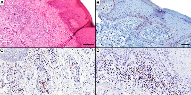
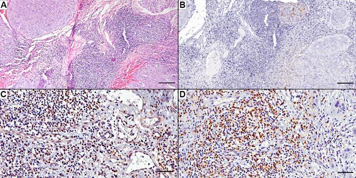
Results: There was a significant difference among the frequencies of CD4+, CD8+, and PD-L1+ cells between AC and LSCC cases, higher in the last group. Moreover, histopathological and atypical changes in AC and LLSCC were correlated with the frequencies of PD-L1+, CD4+, and CD8+ cells. In AC, PD-L1+ cases had a low frequency of CD4+ cells, but on the other hand, PD-L1+ cases of LLSCC had a higher frequency of CD4+ and CD8+ cells.
Conclusion: Therefore, the PD-L1 molecule may be a potential escape route for the immune response in oral lesions, but the mechanisms differ between AC and LLSCC. Future studies related to immune evasion and immunotherapy in oral lesions should consider the analysis of inflammatory infiltrate and TILs.

 求助内容:
求助内容: 应助结果提醒方式:


