Oscar Otero-Marquez, Mona Fayad, Alexander Pinhas, Toco Y P Chui, Richard B Rosen, Harsha S Reddy
{"title":"Retinal Surface Macrophage Changes in Thyroid Eye Disease before and after Treatment with Teprotumumab.","authors":"Oscar Otero-Marquez, Mona Fayad, Alexander Pinhas, Toco Y P Chui, Richard B Rosen, Harsha S Reddy","doi":"10.1155/2022/5275309","DOIUrl":null,"url":null,"abstract":"<p><p>Retinal surface macrophages play key roles in the regulation of immune response, maintenance of vitreous clarity, and tissue repair. We examined the variation of parafoveal surface macrophages in a thyroid eye disease (TED) patient before and after treatment with teprotumumab (Tepezza, Horizon therapeutics). Pre- and posttreatment parafoveal surface macrophages were imaged using clinical <i>en face</i> OCT, and their density was assessed using a novel cell density mapping technique. Pretreatment, surface macrophage cell density was high. Macrophages had a nonuniform spatial distribution, and their appearance was round with few protrusions, consistent with an \"activated\" state. Posttreatment, cell density decreased. The macrophages were regularly spaced and had a ramified appearance and filopodia-like processes, consistent with a \"quiescent\" state. Surface macrophage density decreased as the Clinical Activity Score (CAS) decreased with teprotumumab treatment, suggesting a potential association of these cells with an underlying intraocular and retinal inflammatory process previously not described in TED.</p>","PeriodicalId":9603,"journal":{"name":"Case Reports in Ophthalmological Medicine","volume":null,"pages":null},"PeriodicalIF":0.7000,"publicationDate":"2022-02-07","publicationTypes":"Journal Article","fieldsOfStudy":null,"isOpenAccess":false,"openAccessPdf":"https://www.ncbi.nlm.nih.gov/pmc/articles/PMC8844343/pdf/","citationCount":"2","resultStr":null,"platform":"Semanticscholar","paperid":null,"PeriodicalName":"Case Reports in Ophthalmological Medicine","FirstCategoryId":"1085","ListUrlMain":"https://doi.org/10.1155/2022/5275309","RegionNum":0,"RegionCategory":null,"ArticlePicture":[],"TitleCN":null,"AbstractTextCN":null,"PMCID":null,"EPubDate":"2022/1/1 0:00:00","PubModel":"eCollection","JCR":"Q4","JCRName":"OPHTHALMOLOGY","Score":null,"Total":0}
引用次数: 2
Abstract
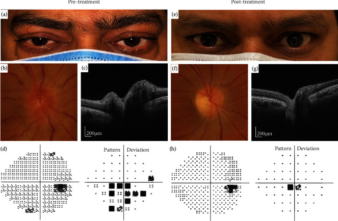
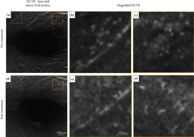
Retinal surface macrophages play key roles in the regulation of immune response, maintenance of vitreous clarity, and tissue repair. We examined the variation of parafoveal surface macrophages in a thyroid eye disease (TED) patient before and after treatment with teprotumumab (Tepezza, Horizon therapeutics). Pre- and posttreatment parafoveal surface macrophages were imaged using clinical en face OCT, and their density was assessed using a novel cell density mapping technique. Pretreatment, surface macrophage cell density was high. Macrophages had a nonuniform spatial distribution, and their appearance was round with few protrusions, consistent with an "activated" state. Posttreatment, cell density decreased. The macrophages were regularly spaced and had a ramified appearance and filopodia-like processes, consistent with a "quiescent" state. Surface macrophage density decreased as the Clinical Activity Score (CAS) decreased with teprotumumab treatment, suggesting a potential association of these cells with an underlying intraocular and retinal inflammatory process previously not described in TED.


 求助内容:
求助内容: 应助结果提醒方式:


