Arosha T. Weerakoon , Crystal Cooper , Ian A. Meyers , Nicholas Condon , Christopher Sexton , David Thomson , Pauline J. Ford , Anne L. Symons
{"title":"Does dentine mineral change with anatomical location, microscopic site and patient age?","authors":"Arosha T. Weerakoon , Crystal Cooper , Ian A. Meyers , Nicholas Condon , Christopher Sexton , David Thomson , Pauline J. Ford , Anne L. Symons","doi":"10.1016/j.yjsbx.2022.100060","DOIUrl":null,"url":null,"abstract":"<div><h3>Objective</h3><p>To determine the effect of patient age (young or mature), anatomical location (shallow/deep and central/peripheral) and microscopic site (intertubular/peritubular) on dentine mineral density, distribution and composition.</p></div><div><h3>Methods</h3><p>Extracted posterior teeth from young (aged 19–20 years, N = 4) and mature (aged 54–77 years, N = 4) subjects were prepared to shallow and deep slices. The dentine surface elemental composition was investigated in a SEM using Backscattered Electron (BSE) micrographs, Energy Dispersive X-ray Spectroscopy, and Integrated Mineral Analysis. Qualitative comparisons and quantitative measures using machine learning were used to analyse the BSE images. Quantitative outcomes were compared using quantile or linear regression models with bootstrapping to account for the multiple measures per sample. Subsequently, a Xenon Plasma Focussed Ion Beam Scanning Electron Microscopy (Xe PFIB-SEM) was used to mill large area (100 µm) cross-sections to investigate morphology through the dentine tubules using high resolution secondary electron micrographs.</p></div><div><h3>Results</h3><p>With age, dentine mineral composition remains stable, but density changes with anatomical location and microscopic site. Microscopically, accessory tubules spread into intertubular dentine (ITD) from the main tubule lumens. Within the lumens, mineral deposits form calcospherites in the young that eventually coalesce in mature tubules and branches. The mineral occlusion in mature dentine increases overall ITD density to reflect peritubular dentine (PTD) infiltrate. The ITD observed in micrographs remained consistent for age and observation plane to suggest tubule deposition affects overall dentine density. Mineral density depends on the relative distribution of PTD to ITD that varies with anatomical location.</p></div><div><h3>Significance</h3><p>Adhesive materials may interact differently within a tooth as well as in different age groups.</p></div>","PeriodicalId":17238,"journal":{"name":"Journal of Structural Biology: X","volume":"6 ","pages":"Article 100060"},"PeriodicalIF":5.1000,"publicationDate":"2022-01-01","publicationTypes":"Journal Article","fieldsOfStudy":null,"isOpenAccess":false,"openAccessPdf":"https://ftp.ncbi.nlm.nih.gov/pub/pmc/oa_pdf/91/16/main.PMC8818708.pdf","citationCount":"4","resultStr":null,"platform":"Semanticscholar","paperid":null,"PeriodicalName":"Journal of Structural Biology: X","FirstCategoryId":"1085","ListUrlMain":"https://www.sciencedirect.com/science/article/pii/S2590152422000010","RegionNum":0,"RegionCategory":null,"ArticlePicture":[],"TitleCN":null,"AbstractTextCN":null,"PMCID":null,"EPubDate":"","PubModel":"","JCR":"Q2","JCRName":"BIOCHEMISTRY & MOLECULAR BIOLOGY","Score":null,"Total":0}
引用次数: 4
Abstract
Objective
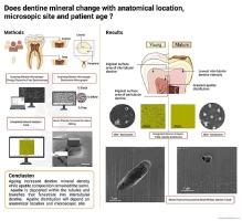
To determine the effect of patient age (young or mature), anatomical location (shallow/deep and central/peripheral) and microscopic site (intertubular/peritubular) on dentine mineral density, distribution and composition.
Methods
Extracted posterior teeth from young (aged 19–20 years, N = 4) and mature (aged 54–77 years, N = 4) subjects were prepared to shallow and deep slices. The dentine surface elemental composition was investigated in a SEM using Backscattered Electron (BSE) micrographs, Energy Dispersive X-ray Spectroscopy, and Integrated Mineral Analysis. Qualitative comparisons and quantitative measures using machine learning were used to analyse the BSE images. Quantitative outcomes were compared using quantile or linear regression models with bootstrapping to account for the multiple measures per sample. Subsequently, a Xenon Plasma Focussed Ion Beam Scanning Electron Microscopy (Xe PFIB-SEM) was used to mill large area (100 µm) cross-sections to investigate morphology through the dentine tubules using high resolution secondary electron micrographs.
Results
With age, dentine mineral composition remains stable, but density changes with anatomical location and microscopic site. Microscopically, accessory tubules spread into intertubular dentine (ITD) from the main tubule lumens. Within the lumens, mineral deposits form calcospherites in the young that eventually coalesce in mature tubules and branches. The mineral occlusion in mature dentine increases overall ITD density to reflect peritubular dentine (PTD) infiltrate. The ITD observed in micrographs remained consistent for age and observation plane to suggest tubule deposition affects overall dentine density. Mineral density depends on the relative distribution of PTD to ITD that varies with anatomical location.
Significance
Adhesive materials may interact differently within a tooth as well as in different age groups.
 求助内容:
求助内容: 应助结果提醒方式:


