Pelsin Demir, Nathaniel Hovsepian, Peter Pagels, Vanja Petersson, Karthikeyan Baskaran, Antonio Filipe Macedo
{"title":"All retinas are not created equal: Fovea-to-macula thickness ratio and foveal microvasculature in healthy young children.","authors":"Pelsin Demir, Nathaniel Hovsepian, Peter Pagels, Vanja Petersson, Karthikeyan Baskaran, Antonio Filipe Macedo","doi":"10.1111/opo.12958","DOIUrl":null,"url":null,"abstract":"<p><strong>Purpose: </strong>Markers for the relationships between structural and microvasculature measures given by optical coherence tomography angiography are necessary to increase the diagnostic and prognostic value of this technique. The aim of this study was to investigate relationships between structural and microvasculature measures around the fovea in healthy eyes of healthy children.</p><p><strong>Methods: </strong>Observational cross-sectional study involving children aged 8-17 years, born at full-term, with no eye disease. The better of two 3 × 3 mm macular scans obtained with a Cirrus 5000HD-OCT was analysed. Images were corrected for lateral magnification errors. Vessel density and perfusion were measured with ImageJ/Fiji software for the superficial capillary plexus. Structural measures including foveal and macular thicknesses were performed manually.</p><p><strong>Results: </strong>The sample included 86 participants, 51 (59%) females. Mean age was 12.4 years (SD = 2.5); mean best-corrected acuity was -0.10 logMAR (SD = 0.09); mean refractive error was +0.59 D (SD = 1.3) and mean axial length was 23.1 mm (SD = 0.86). Mean area of the foveal avascular zone (AFAZ) was 0.20 mm<sup>2</sup> (SD = 0.88); median fovea-to-macula thickness ratio (FMTR) was 0.63 (IQR = 0.08); mean central vessel density was 12.42 mm<sup>-1</sup> (SD = 2.78) and mean central perfusion was 38.66% (SD = 3.83). AFAZ was correlated with central vessel density (p < 0.001), perfusion (p < 0.001), foveal thickness (p < 0.001) and FMTR (p < 0.001). Central vessel density was correlated with foveal thickness (p < 0.001) and FMTR, (p = 0.01). Central perfusion was correlated with foveal thickness (p < 0.001) and FMTR, (p = 0.003).</p><p><strong>Conclusion: </strong>In this study, foveal thickness, FMTR and foveal microvasculature measurements were correlated. Clinicians need to be aware that shallow foveal pits and persistent foveal microvasculature are likely to occur in optical coherence tomography angiography images. In healthy eyes from healthy children, an atypical high FMTR and a small AFAZ may be associated with incomplete foveal development. The mechanism and functional implications of this remain unknown.</p>","PeriodicalId":520731,"journal":{"name":"Ophthalmic & physiological optics : the journal of the British College of Ophthalmic Opticians (Optometrists)","volume":" ","pages":"644-652"},"PeriodicalIF":2.4000,"publicationDate":"2022-05-01","publicationTypes":"Journal Article","fieldsOfStudy":null,"isOpenAccess":false,"openAccessPdf":"https://ftp.ncbi.nlm.nih.gov/pub/pmc/oa_pdf/3d/b6/OPO-42-644.PMC9304185.pdf","citationCount":"1","resultStr":null,"platform":"Semanticscholar","paperid":null,"PeriodicalName":"Ophthalmic & physiological optics : the journal of the British College of Ophthalmic Opticians (Optometrists)","FirstCategoryId":"3","ListUrlMain":"https://doi.org/10.1111/opo.12958","RegionNum":0,"RegionCategory":null,"ArticlePicture":[],"TitleCN":null,"AbstractTextCN":null,"PMCID":null,"EPubDate":"2022/2/14 0:00:00","PubModel":"Epub","JCR":"","JCRName":"","Score":null,"Total":0}
引用次数: 1
Abstract
Purpose: Markers for the relationships between structural and microvasculature measures given by optical coherence tomography angiography are necessary to increase the diagnostic and prognostic value of this technique. The aim of this study was to investigate relationships between structural and microvasculature measures around the fovea in healthy eyes of healthy children.
Methods: Observational cross-sectional study involving children aged 8-17 years, born at full-term, with no eye disease. The better of two 3 × 3 mm macular scans obtained with a Cirrus 5000HD-OCT was analysed. Images were corrected for lateral magnification errors. Vessel density and perfusion were measured with ImageJ/Fiji software for the superficial capillary plexus. Structural measures including foveal and macular thicknesses were performed manually.
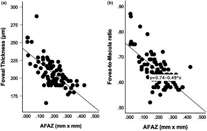
Results: The sample included 86 participants, 51 (59%) females. Mean age was 12.4 years (SD = 2.5); mean best-corrected acuity was -0.10 logMAR (SD = 0.09); mean refractive error was +0.59 D (SD = 1.3) and mean axial length was 23.1 mm (SD = 0.86). Mean area of the foveal avascular zone (AFAZ) was 0.20 mm2 (SD = 0.88); median fovea-to-macula thickness ratio (FMTR) was 0.63 (IQR = 0.08); mean central vessel density was 12.42 mm-1 (SD = 2.78) and mean central perfusion was 38.66% (SD = 3.83). AFAZ was correlated with central vessel density (p < 0.001), perfusion (p < 0.001), foveal thickness (p < 0.001) and FMTR (p < 0.001). Central vessel density was correlated with foveal thickness (p < 0.001) and FMTR, (p = 0.01). Central perfusion was correlated with foveal thickness (p < 0.001) and FMTR, (p = 0.003).
Conclusion: In this study, foveal thickness, FMTR and foveal microvasculature measurements were correlated. Clinicians need to be aware that shallow foveal pits and persistent foveal microvasculature are likely to occur in optical coherence tomography angiography images. In healthy eyes from healthy children, an atypical high FMTR and a small AFAZ may be associated with incomplete foveal development. The mechanism and functional implications of this remain unknown.

 求助内容:
求助内容: 应助结果提醒方式:


