Hanna Hebelka, Mohammad Khalil, Helena Brisby, Kerstin Lagerstrand
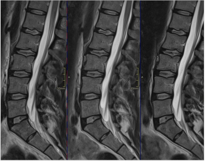
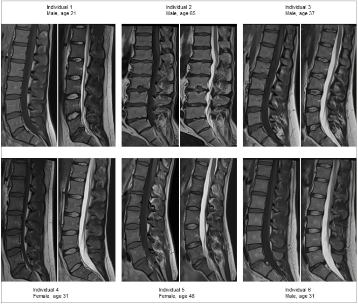
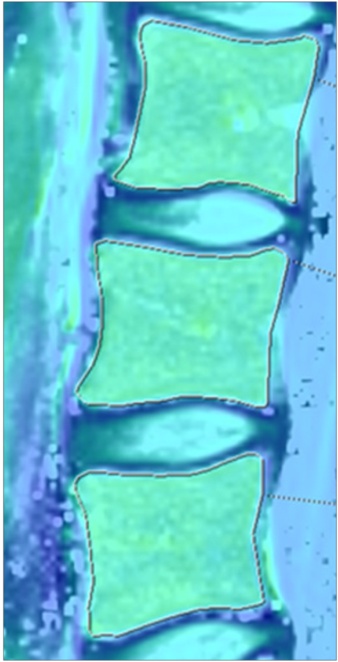
{"title":"Lumbar vertebral T2-relaxation time investigated with T2-mapping at multiple time points in a day demonstrate large individual variations.","authors":"Hanna Hebelka, Mohammad Khalil, Helena Brisby, Kerstin Lagerstrand","doi":"10.5152/dir.2021.21514","DOIUrl":null,"url":null,"abstract":"<p><p>PURPOSE The increasing interest of endplate and Modic changes as potential pain generators in low back pain (LBP), along with advancement of functional quantitative magnetic resonance imaging (MRI) techniques, makes it important to characterize the vertebral dynamic behavior in detail. This study aims to perform characterization of the dynamic behavior of the vertebral bodies (VB) by investigating the VB diurnal variation in T2-relaxation time in a cross-sectional asymptomatic group of individuals. METHODS T2-mapping of 30 VBs (L1-L5) in six healthy volunteers (mean age, 40 years; range, 29-65 years) was performed with a 1.5 Tesla MRI at three time points over the day (7 am, 12 am, 5 pm). Volumetric regions of interest were segmented manually to determine VB T2-relaxation time, which was compared between the three time points. RESULTS On a group level only small and not significant diurnal VB variation was detected (all P >.10), with median T2 (ms) (quartiles; Q1, Q3) at the three time points 88.7 (84.1, 99.1), 87.3 (85.0, 96.1) and 87.8 (84.4, 99.2). However, in some VBs up to 7% increase respectively 9% decrease in T2-relaxation time was found during the day. Further, there was a relatively large variation between the individuals in absolute VB T2-relaxation times (range 73.2-108.3 ms), but small differences between the VBs within an individual. CONCLUSION This first T2-mapping study of the VB signal dynamics, in repeated investigations during one day, display variation in T2-relaxation time in specific individual VBs but were negligible on a group level. The result may be of importance when evaluating patients with spinal pathologies and suggest further examinations of dynamic changes not only of the disc but also vertebrae.</p>","PeriodicalId":50582,"journal":{"name":"Diagnostic and Interventional Radiology","volume":" ","pages":"92-97"},"PeriodicalIF":1.7000,"publicationDate":"2022-01-01","publicationTypes":"Journal Article","fieldsOfStudy":null,"isOpenAccess":false,"openAccessPdf":"https://www.ncbi.nlm.nih.gov/pmc/articles/PMC12278926/pdf/","citationCount":"0","resultStr":null,"platform":"Semanticscholar","paperid":null,"PeriodicalName":"Diagnostic and Interventional Radiology","FirstCategoryId":"3","ListUrlMain":"https://doi.org/10.5152/dir.2021.21514","RegionNum":4,"RegionCategory":"医学","ArticlePicture":[],"TitleCN":null,"AbstractTextCN":null,"PMCID":null,"EPubDate":"","PubModel":"","JCR":"Q2","JCRName":"Medicine","Score":null,"Total":0}
引用次数: 0
Abstract
PURPOSE The increasing interest of endplate and Modic changes as potential pain generators in low back pain (LBP), along with advancement of functional quantitative magnetic resonance imaging (MRI) techniques, makes it important to characterize the vertebral dynamic behavior in detail. This study aims to perform characterization of the dynamic behavior of the vertebral bodies (VB) by investigating the VB diurnal variation in T2-relaxation time in a cross-sectional asymptomatic group of individuals. METHODS T2-mapping of 30 VBs (L1-L5) in six healthy volunteers (mean age, 40 years; range, 29-65 years) was performed with a 1.5 Tesla MRI at three time points over the day (7 am, 12 am, 5 pm). Volumetric regions of interest were segmented manually to determine VB T2-relaxation time, which was compared between the three time points. RESULTS On a group level only small and not significant diurnal VB variation was detected (all P >.10), with median T2 (ms) (quartiles; Q1, Q3) at the three time points 88.7 (84.1, 99.1), 87.3 (85.0, 96.1) and 87.8 (84.4, 99.2). However, in some VBs up to 7% increase respectively 9% decrease in T2-relaxation time was found during the day. Further, there was a relatively large variation between the individuals in absolute VB T2-relaxation times (range 73.2-108.3 ms), but small differences between the VBs within an individual. CONCLUSION This first T2-mapping study of the VB signal dynamics, in repeated investigations during one day, display variation in T2-relaxation time in specific individual VBs but were negligible on a group level. The result may be of importance when evaluating patients with spinal pathologies and suggest further examinations of dynamic changes not only of the disc but also vertebrae.
期刊介绍:
Diagnostic and Interventional Radiology (Diagn Interv Radiol) is the open access, online-only official publication of Turkish Society of Radiology. It is published bimonthly and the journal’s publication language is English.
The journal is a medium for original articles, reviews, pictorial essays, technical notes related to all fields of diagnostic and interventional radiology.

 求助内容:
求助内容: 应助结果提醒方式:


