Where is my ear? - Cervical Chondrocutaneous Branchial Remnant.

IF 0.7 Q4 OTORHINOLARYNGOLOGY
Turkish Archives of Otorhinolaryngology Pub Date : 2021-09-01 Epub Date: 2021-10-15 DOI:10.4274/tao.2021.2021-6-13
Smile Kajal, Anam Ahmed, Anurag Gupta
{"title":"Where is my ear? - Cervical Chondrocutaneous Branchial Remnant.","authors":"Smile Kajal, Anam Ahmed, Anurag Gupta","doi":"10.4274/tao.2021.2021-6-13","DOIUrl":null,"url":null,"abstract":"A 6-year-old boy presented with the absence of pinna on the left side since birth. A cutaneous appendage emerging from the neck on the left side was discovered during the examination (Figure 1). Deep palpation revealed a firm texture like cartilage. On the left side, a primitive firm cartilage was palpated in the helix region (Figure 2). The rest of the cartilaginous framework and external auditory canal (EAC) opening was absent on the left side. On the right side, the pinna was smaller in size and loped. High resolution computed tomography (HRCT) of temporal bone showed an atretic and stenosed EAC on the left and ride side, respectively. Ossicular lump was noted on both sides on HRCT, but all inner ear structures appeared normal in both ears. Pure tone audiometry revealed moderate and mild conductive hearing loss on the left and right sides, respectively. Magnetic resonance imaging of the neck was done to rule out any branchial abnormality. It did not reveal any cyst/sinus/fistula. A provisional diagnosis of cervical chondrocutaneous branchial remnant (CCBR) with leftsided microtia and EAC atresia was made on clinico-radiological basis. After a thorough discussion with the father, the patient was planned for the excision of the cervical mass and staged pinnaplasty and canaloplasty on the left side at a later stage. Histopathological examination after excision showed elastic cartilage rests covered by normal skin consisting of epidermis, dermis, adnexal structures, and subcutaneous fat compatible with CCBR.","PeriodicalId":44240,"journal":{"name":"Turkish Archives of Otorhinolaryngology","volume":"59 3","pages":"242-243"},"PeriodicalIF":0.7000,"publicationDate":"2021-09-01","publicationTypes":"Journal Article","fieldsOfStudy":null,"isOpenAccess":false,"openAccessPdf":"https://ftp.ncbi.nlm.nih.gov/pub/pmc/oa_pdf/94/19/tao-59-242.PMC8527535.pdf","citationCount":"2","resultStr":null,"platform":"Semanticscholar","paperid":null,"PeriodicalName":"Turkish Archives of Otorhinolaryngology","FirstCategoryId":"1085","ListUrlMain":"https://doi.org/10.4274/tao.2021.2021-6-13","RegionNum":0,"RegionCategory":null,"ArticlePicture":[],"TitleCN":null,"AbstractTextCN":null,"PMCID":null,"EPubDate":"2021/10/15 0:00:00","PubModel":"Epub","JCR":"Q4","JCRName":"OTORHINOLARYNGOLOGY","Score":null,"Total":0}
引用次数: 2

Abstract

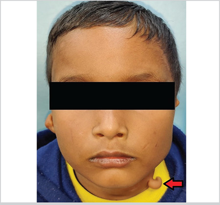
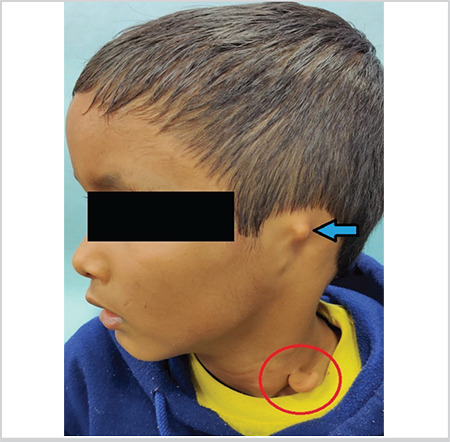
A 6-year-old boy presented with the absence of pinna on the left side since birth. A cutaneous appendage emerging from the neck on the left side was discovered during the examination (Figure 1). Deep palpation revealed a firm texture like cartilage. On the left side, a primitive firm cartilage was palpated in the helix region (Figure 2). The rest of the cartilaginous framework and external auditory canal (EAC) opening was absent on the left side. On the right side, the pinna was smaller in size and loped. High resolution computed tomography (HRCT) of temporal bone showed an atretic and stenosed EAC on the left and ride side, respectively. Ossicular lump was noted on both sides on HRCT, but all inner ear structures appeared normal in both ears. Pure tone audiometry revealed moderate and mild conductive hearing loss on the left and right sides, respectively. Magnetic resonance imaging of the neck was done to rule out any branchial abnormality. It did not reveal any cyst/sinus/fistula. A provisional diagnosis of cervical chondrocutaneous branchial remnant (CCBR) with leftsided microtia and EAC atresia was made on clinico-radiological basis. After a thorough discussion with the father, the patient was planned for the excision of the cervical mass and staged pinnaplasty and canaloplasty on the left side at a later stage. Histopathological examination after excision showed elastic cartilage rests covered by normal skin consisting of epidermis, dermis, adnexal structures, and subcutaneous fat compatible with CCBR.
Abstract Image

Abstract Image

我的耳朵在哪里?-颈椎软骨皮鳃裂残余。
本文章由计算机程序翻译,如有差异,请以英文原文为准。
求助全文
约1分钟内获得全文 求助全文
来源期刊
自引率
0.00%
发文量
0
×
引用
GB/T 7714-2015
复制
MLA
复制
APA
复制
导出至
BibTeX EndNote RefMan NoteFirst NoteExpress
×
提示
您的信息不完整,为了账户安全,请先补充。
现在去补充
×
提示
您因"违规操作"
具体请查看互助需知
我知道了
×
提示
确定
请完成安全验证×
copy
已复制链接
快去分享给好友吧!
我知道了
右上角分享
点击右上角分享
0
联系我们:info@booksci.cn Book学术提供免费学术资源搜索服务,方便国内外学者检索中英文文献。致力于提供最便捷和优质的服务体验。 Copyright © 2023 布克学术 All rights reserved.
京ICP备2023020795号-1
ghs 京公网安备 11010802042870号
Book学术文献互助
Book学术文献互助群
群 号:481959085
Book学术官方微信