Jeonghwan Lee, Jung-Woo Son, Siekyeong Kim, Ji-Eun Kim, Seungwon Chung, Hei-Rhee Ghim, Sang-Ick Lee, Chul-Jin Shin, Gawon Ju
{"title":"Disrupted Association Between Empathy and Brain Structure in Attention-Deficit/Hyperactivity Disorder.","authors":"Jeonghwan Lee, Jung-Woo Son, Siekyeong Kim, Ji-Eun Kim, Seungwon Chung, Hei-Rhee Ghim, Sang-Ick Lee, Chul-Jin Shin, Gawon Ju","doi":"10.5765/jkacap.210009","DOIUrl":null,"url":null,"abstract":"<p><strong>Objectives: </strong>To investigate the relationship between brain structure and empathy in early adolescents with attention-deficit/hyperactivity disorder (ADHD).</p><p><strong>Methods: </strong>Nineteen early adolescents with ADHD and 20 healthy controls underwent 3T MRI. All the participants were assessed for different aspects of empathy using measures including the Interpersonal Reactivity Index and Empathy Quotient. Cortical thickness and subcortical structural volume based on T1-weighted scans were analyzed using FreeSurfer.</p><p><strong>Results: </strong>Cognitive empathy (t=-2.52, p=0.016) and perspective taking (t=-2.10, p=0.043) were impaired in the ADHD group compared with the control group. The cluster encompassing the left posterior insular, supramarginal, and transverse temporal cortices [cluster-wise p-value (CWP)=0.001], which are associated with emotional empathy, was significantly smaller in the ADHD group, and the volume of the left nucleus accumbens was greater than that of the control group (F=10.12, p=0.003, effect size=0.22). In the control group, the left superior temporal (CWP=0.002) and lingual cortical (CWP=0.035) thicknesses were positively associated with cognitive empathy, while the right amygdala volume was positively associated with empathic concern (Coef=14.26, t=3.92, p=0.001). However, there was no significant correlation between empathy and brain structure in the ADHD group.</p><p><strong>Conclusion: </strong>The ADHD group had a smaller volume of the cortical area associated with emotional empathy than the control group, and there was no brain region showing significant correlation with empathy, unlike in the control group.</p>","PeriodicalId":42806,"journal":{"name":"Journal of the Korean Academy of Child and Adolescent Psychiatry","volume":"32 4","pages":"129-136"},"PeriodicalIF":1.4000,"publicationDate":"2021-10-01","publicationTypes":"Journal Article","fieldsOfStudy":null,"isOpenAccess":false,"openAccessPdf":"https://ftp.ncbi.nlm.nih.gov/pub/pmc/oa_pdf/88/d3/jkacap-32-4-129.PMC8499037.pdf","citationCount":"1","resultStr":null,"platform":"Semanticscholar","paperid":null,"PeriodicalName":"Journal of the Korean Academy of Child and Adolescent Psychiatry","FirstCategoryId":"1085","ListUrlMain":"https://doi.org/10.5765/jkacap.210009","RegionNum":0,"RegionCategory":null,"ArticlePicture":[],"TitleCN":null,"AbstractTextCN":null,"PMCID":null,"EPubDate":"","PubModel":"","JCR":"Q4","JCRName":"PSYCHIATRY","Score":null,"Total":0}
引用次数: 1
Abstract
Objectives: To investigate the relationship between brain structure and empathy in early adolescents with attention-deficit/hyperactivity disorder (ADHD).
Methods: Nineteen early adolescents with ADHD and 20 healthy controls underwent 3T MRI. All the participants were assessed for different aspects of empathy using measures including the Interpersonal Reactivity Index and Empathy Quotient. Cortical thickness and subcortical structural volume based on T1-weighted scans were analyzed using FreeSurfer.
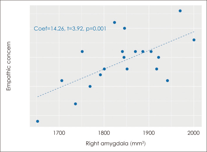
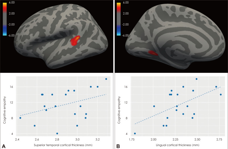
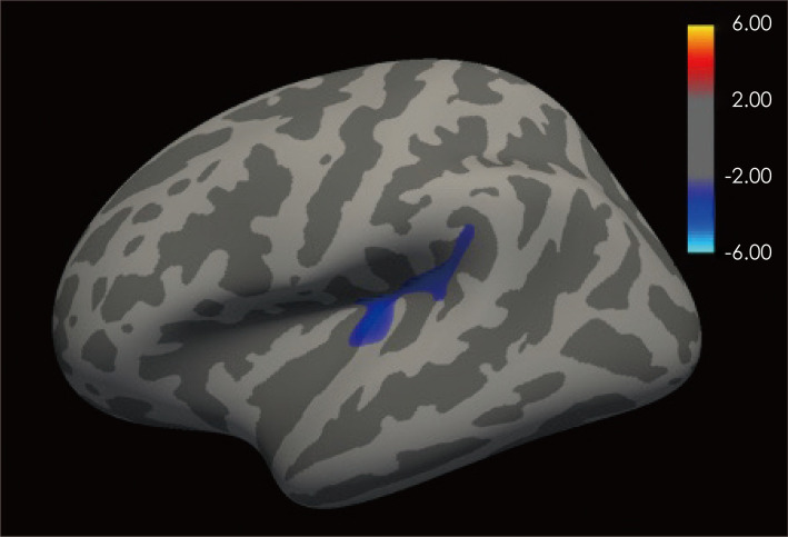
Results: Cognitive empathy (t=-2.52, p=0.016) and perspective taking (t=-2.10, p=0.043) were impaired in the ADHD group compared with the control group. The cluster encompassing the left posterior insular, supramarginal, and transverse temporal cortices [cluster-wise p-value (CWP)=0.001], which are associated with emotional empathy, was significantly smaller in the ADHD group, and the volume of the left nucleus accumbens was greater than that of the control group (F=10.12, p=0.003, effect size=0.22). In the control group, the left superior temporal (CWP=0.002) and lingual cortical (CWP=0.035) thicknesses were positively associated with cognitive empathy, while the right amygdala volume was positively associated with empathic concern (Coef=14.26, t=3.92, p=0.001). However, there was no significant correlation between empathy and brain structure in the ADHD group.
Conclusion: The ADHD group had a smaller volume of the cortical area associated with emotional empathy than the control group, and there was no brain region showing significant correlation with empathy, unlike in the control group.

 求助内容:
求助内容: 应助结果提醒方式:


