Optical Coherence Tomography Angiography of Early Stage 1a Retinal Hemangioblastoma in Von-Hippel-Lindau.
IF 1.9
Q3 ONCOLOGY
Journal of Kidney Cancer and VHL
Pub Date : 2021-09-23
eCollection Date: 2021-01-01
DOI:10.15586/jkcvhl.v8i3.158
引用次数: 1
Abstract
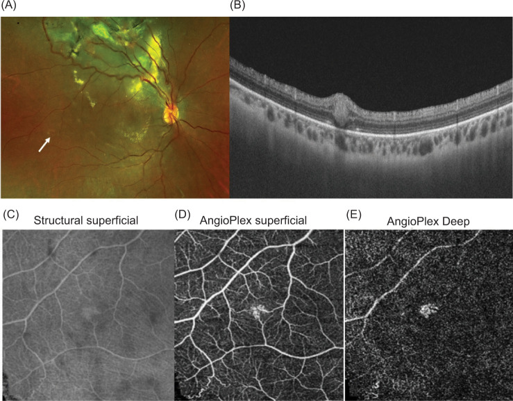
Von-Hippel-Lindau (VHL) syndrome is characterized by focal vasoproliferative tumors of retinal capillaries called retinal capillary hemangioblastomas (RCH). These tumors are initially small and can be easily missed if not looked for carefully. As they grow, these tumors are more demanding to treat and hence the importance of detecting them early and treating them. Herein, we describe and review the optical coherence tomography angiography (OCTA) of the early- stage lesion, which suggested the involvement of superficial and a deeper retinal capillary plexus. In addition, to helping us detect these lesions earlier, OCTA may also help to understand the in vivo changes occurring at an earlier phase.

Von-Hippel-Lindau早期1a期视网膜血管母细胞瘤的光学相干断层血管造影。
Von-Hippel-Lindau (VHL)综合征的特征是视网膜毛细血管的局灶性血管增生性肿瘤,称为视网膜毛细血管母细胞瘤(RCH)。这些肿瘤最初很小,如果不仔细检查很容易被遗漏。随着肿瘤的生长,治疗难度越来越大,因此及早发现和治疗非常重要。在此,我们描述并回顾了光学相干断层血管造影(OCTA)的早期病变,提示浅层和深层视网膜毛细血管丛受累。此外,为了帮助我们更早地发现这些病变,OCTA也可能有助于了解发生在早期阶段的体内变化。
本文章由计算机程序翻译,如有差异,请以英文原文为准。
求助全文
约1分钟内获得全文
求助全文

 求助内容:
求助内容: 应助结果提醒方式:


