Etienne Côté, Rong-Mo Zhang, Nicole Kaiser, Dieter P Reinhardt, Chelsea K Martin
{"title":"Annuloaortic ectasia in a four-month-old male Newfoundland dog: long-term follow-up and immunofluorescent study.","authors":"Etienne Côté, Rong-Mo Zhang, Nicole Kaiser, Dieter P Reinhardt, Chelsea K Martin","doi":"10.1080/01652176.2021.1961039","DOIUrl":null,"url":null,"abstract":"<p><p>A 4 month-old, 14.8 kg, male Newfoundland dog was presented for cardiovascular evaluation following detection of a heart murmur. Echocardiography revealed enlargement of the sinuses of Valsalva and marked, diffuse dilation of the ascending aorta (annuloaortic ectasia, AAE), with mild/equivocal subaortic stenosis (SAS). The dog was monitored over the duration of its lifetime, with serial echocardiograms performed at 5, 6, and 8 months and 1, 2, 3, 4, 8, and 10 years demonstrating persistent, diffuse dilation of the ascending aorta. The dog lived until it was 10 years old and died of metastatic carcinoma. Postmortem examination confirmed AAE and mild SAS. Hematoxylin and eosin and Weigert van Gieson stains were used to compare the ascending aorta to the descending aorta and left subclavian artery, and to compare aortic samples to those of three control dogs. Histopathologic evaluation revealed mild medial degeneration in the ascending aorta of all four dogs. Immunofluorescent microscopy was used for determining the deposition of proteins known to play a role in aortic aneurysms in humans: fibrillin-1 (FBN1), latent transforming growth factor beta binding protein 4 (LTBP4) and fibronectin. The ascending aorta of the AAE case demonstrated reduced deposition of FBN1, indicating that its loss may have contributed to aortic dilation. Diffuse, primary ascending aortic dilation is uncommonly reported in dogs; when it is, it carries a poor prognosis. This case provides an important example of marked dilation of the ascending aorta in a dog that lived with no associated adverse effects for 10 years.</p>","PeriodicalId":51207,"journal":{"name":"Veterinary Quarterly","volume":null,"pages":null},"PeriodicalIF":7.9000,"publicationDate":"2021-12-01","publicationTypes":"Journal Article","fieldsOfStudy":null,"isOpenAccess":false,"openAccessPdf":"https://www.ncbi.nlm.nih.gov/pmc/articles/PMC8526017/pdf/","citationCount":"0","resultStr":null,"platform":"Semanticscholar","paperid":null,"PeriodicalName":"Veterinary Quarterly","FirstCategoryId":"97","ListUrlMain":"https://doi.org/10.1080/01652176.2021.1961039","RegionNum":2,"RegionCategory":"农林科学","ArticlePicture":[],"TitleCN":null,"AbstractTextCN":null,"PMCID":null,"EPubDate":"","PubModel":"","JCR":"Q1","JCRName":"VETERINARY SCIENCES","Score":null,"Total":0}
引用次数: 0
Abstract
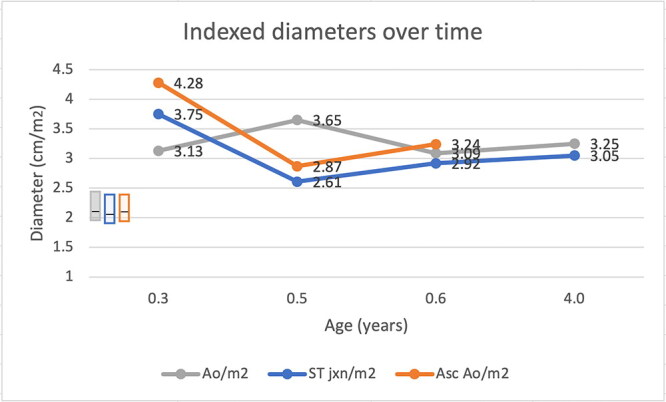
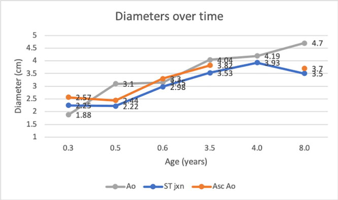
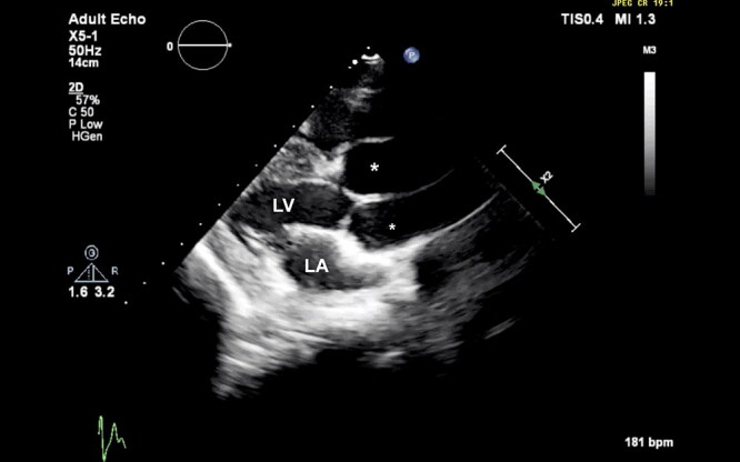
A 4 month-old, 14.8 kg, male Newfoundland dog was presented for cardiovascular evaluation following detection of a heart murmur. Echocardiography revealed enlargement of the sinuses of Valsalva and marked, diffuse dilation of the ascending aorta (annuloaortic ectasia, AAE), with mild/equivocal subaortic stenosis (SAS). The dog was monitored over the duration of its lifetime, with serial echocardiograms performed at 5, 6, and 8 months and 1, 2, 3, 4, 8, and 10 years demonstrating persistent, diffuse dilation of the ascending aorta. The dog lived until it was 10 years old and died of metastatic carcinoma. Postmortem examination confirmed AAE and mild SAS. Hematoxylin and eosin and Weigert van Gieson stains were used to compare the ascending aorta to the descending aorta and left subclavian artery, and to compare aortic samples to those of three control dogs. Histopathologic evaluation revealed mild medial degeneration in the ascending aorta of all four dogs. Immunofluorescent microscopy was used for determining the deposition of proteins known to play a role in aortic aneurysms in humans: fibrillin-1 (FBN1), latent transforming growth factor beta binding protein 4 (LTBP4) and fibronectin. The ascending aorta of the AAE case demonstrated reduced deposition of FBN1, indicating that its loss may have contributed to aortic dilation. Diffuse, primary ascending aortic dilation is uncommonly reported in dogs; when it is, it carries a poor prognosis. This case provides an important example of marked dilation of the ascending aorta in a dog that lived with no associated adverse effects for 10 years.
一只4个月大,14.8公斤的雄性纽芬兰犬在检测到心脏杂音后进行了心血管评估。超声心动图显示Valsalva窦增大,升主动脉明显弥漫性扩张(主动脉环扩张,AAE),伴轻度/模棱两可的主动脉下狭窄(SAS)。在狗的一生中进行监测,在5、6和8个月以及1、2、3、4、8和10年进行连续超声心动图检查,显示持续的、弥漫性的升主动脉扩张。这只狗活到了10岁,死于转移性癌。尸检证实为AAE和轻度SAS。采用苏木精、伊红染色和Weigert van Gieson染色比较升主动脉、降主动脉和左锁骨下动脉,并将主动脉样本与三只对照犬的主动脉样本进行比较。组织病理学评估显示,所有4只狗的升主动脉均有轻度内侧变性。免疫荧光显微镜用于测定已知在人主动脉瘤中起作用的蛋白的沉积:纤维蛋白-1 (FBN1)、潜伏转化生长因子β结合蛋白4 (LTBP4)和纤维连接蛋白。AAE病例的升主动脉显示FBN1沉积减少,表明其丢失可能导致主动脉扩张。弥漫性,原发性升主动脉扩张是罕见的报道在狗;一旦发生,预后就很差。这个病例提供了一个重要的例子,在一个狗的显著扩张的升主动脉,没有相关的不良反应生活了10年。
期刊介绍:
Veterinary Quarterly is an international open access journal which publishes high quality review articles and original research in the field of veterinary science and animal diseases. The journal publishes research on a range of different animal species and topics including: - Economically important species such as domesticated and non-domesticated farm animals, including avian and poultry diseases; - Companion animals (dogs, cats, horses, pocket pets and exotics); - Wildlife species; - Infectious diseases; - Diagnosis; - Treatment including pharmacology and vaccination
 求助内容:
求助内容: 应助结果提醒方式:


