{"title":"Quantitative Susceptibility Mapping versus R2*-based Histogram Analysis for Evaluating Liver Fibrosis: Preliminary Results.","authors":"Masato Yoshikawa, Kohsuke Kudo, Taisuke Harada, Kazutaka Harashima, Jun Suzuki, Koji Ogawa, Taro Fujiwara, Mutsumi Nishida, Ryota Sato, Toru Shirai, Yoshitaka Bito","doi":"10.2463/mrms.mp.2020-0175","DOIUrl":null,"url":null,"abstract":"<p><strong>Purpose: </strong>The staging of liver fibrosis is clinically important, and a less invasive method is preferred. Quantitative susceptibility mapping (QSM) has shown a great potential in estimating liver fibrosis in addition to R2* relaxometry. However, few studies have compared QSM analysis and liver fibrosis. We aimed to evaluate the feasibility of estimating liver fibrosis by using QSM and R2*-based histogram analyses by comparing it with ultrasound-based transient elastography and the stage of histologic fibrosis.</p><p><strong>Methods: </strong>Fourteen patients with liver disease were enrolled. Data sets of multi-echo gradient echo sequence with breath-holding were acquired on a 3-Tesla scanner. QSM and R2* were reconstructed by water-fat separation method, and ROIs were analyzed for these images. Quantitative parameters with histogram features (mean, variance, skewness, kurtosis, and 1st, 10th, 50th, 90th, and 99th percentiles) were extracted. These data were compared with the elasticity measured by ultrasound transient elastography and histological stage of liver fibrosis (F0 to F4, based on the new Inuyama classification) determined by biopsy or hepatectomy. The correlation of histogram parameters with intrahepatic elasticity and histologically confirmed fibrosis stage was examined. Texture parameters were compared between subgroups divided according to fibrosis stage. Receiver operating characteristic (ROC) analysis was also performed. P < 0.05 indicated statistical significance.</p><p><strong>Results: </strong>The six histogram parameters of both QSM and R2*were significantly correlated with intrahepatic elasticity. In particular, three parameters (variance, percentiles [90th and 99th]) of QSM showed high correlation (r = 0.818-0.844), whereas R2* parameters showed a moderate correlation with elasticity. Four parameters of QSM were significantly correlated with fibrosis stage (ρ = 0.637-0.723) and differentiated F2-4 from F0-1 fibrosis and F3-4 from F0-2 fibrosis with areas under the ROC curve of > 0.8, but those of R2* did not.</p><p><strong>Conclusion: </strong>QSM may serve as a promising surrogate indicator in detecting liver fibrosis.</p>","PeriodicalId":18119,"journal":{"name":"Magnetic Resonance in Medical Sciences","volume":"21 4","pages":"609-622"},"PeriodicalIF":3.2000,"publicationDate":"2022-10-01","publicationTypes":"Journal Article","fieldsOfStudy":null,"isOpenAccess":false,"openAccessPdf":"https://ftp.ncbi.nlm.nih.gov/pub/pmc/oa_pdf/60/91/mrms-21-609.PMC9618931.pdf","citationCount":"4","resultStr":null,"platform":"Semanticscholar","paperid":null,"PeriodicalName":"Magnetic Resonance in Medical Sciences","FirstCategoryId":"3","ListUrlMain":"https://doi.org/10.2463/mrms.mp.2020-0175","RegionNum":3,"RegionCategory":"医学","ArticlePicture":[],"TitleCN":null,"AbstractTextCN":null,"PMCID":null,"EPubDate":"2021/11/11 0:00:00","PubModel":"Epub","JCR":"Q2","JCRName":"RADIOLOGY, NUCLEAR MEDICINE & MEDICAL IMAGING","Score":null,"Total":0}
引用次数: 4
Abstract
Purpose: The staging of liver fibrosis is clinically important, and a less invasive method is preferred. Quantitative susceptibility mapping (QSM) has shown a great potential in estimating liver fibrosis in addition to R2* relaxometry. However, few studies have compared QSM analysis and liver fibrosis. We aimed to evaluate the feasibility of estimating liver fibrosis by using QSM and R2*-based histogram analyses by comparing it with ultrasound-based transient elastography and the stage of histologic fibrosis.
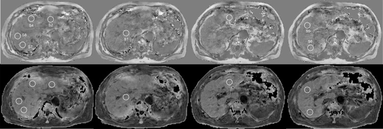
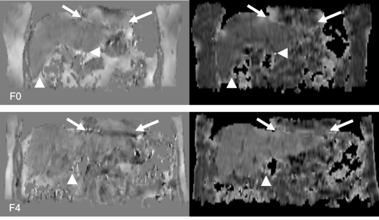
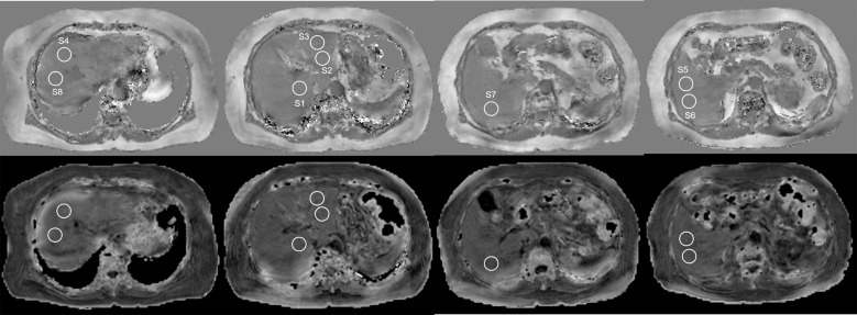
Methods: Fourteen patients with liver disease were enrolled. Data sets of multi-echo gradient echo sequence with breath-holding were acquired on a 3-Tesla scanner. QSM and R2* were reconstructed by water-fat separation method, and ROIs were analyzed for these images. Quantitative parameters with histogram features (mean, variance, skewness, kurtosis, and 1st, 10th, 50th, 90th, and 99th percentiles) were extracted. These data were compared with the elasticity measured by ultrasound transient elastography and histological stage of liver fibrosis (F0 to F4, based on the new Inuyama classification) determined by biopsy or hepatectomy. The correlation of histogram parameters with intrahepatic elasticity and histologically confirmed fibrosis stage was examined. Texture parameters were compared between subgroups divided according to fibrosis stage. Receiver operating characteristic (ROC) analysis was also performed. P < 0.05 indicated statistical significance.
Results: The six histogram parameters of both QSM and R2*were significantly correlated with intrahepatic elasticity. In particular, three parameters (variance, percentiles [90th and 99th]) of QSM showed high correlation (r = 0.818-0.844), whereas R2* parameters showed a moderate correlation with elasticity. Four parameters of QSM were significantly correlated with fibrosis stage (ρ = 0.637-0.723) and differentiated F2-4 from F0-1 fibrosis and F3-4 from F0-2 fibrosis with areas under the ROC curve of > 0.8, but those of R2* did not.
Conclusion: QSM may serve as a promising surrogate indicator in detecting liver fibrosis.
期刊介绍:
Magnetic Resonance in Medical Sciences (MRMS or Magn
Reson Med Sci) is an international journal pursuing the
publication of original articles contributing to the progress
of magnetic resonance in the field of biomedical sciences
including technical developments and clinical applications.
MRMS is an official journal of the Japanese Society for
Magnetic Resonance in Medicine (JSMRM).
 求助内容:
求助内容: 应助结果提醒方式:


