The Evaluation of Tau Deposition with [18F]PI-2620 by Using a Semiquantitative Method in Cognitively Normal Subjects and Patients with Mild Cognitive Impairment and Alzheimer's Disease.
{"title":"The Evaluation of Tau Deposition with [<sup>18</sup>F]PI-2620 by Using a Semiquantitative Method in Cognitively Normal Subjects and Patients with Mild Cognitive Impairment and Alzheimer's Disease.","authors":"Attapon Jantarato, Sira Vachatimanont, Natphimol Boonkawin, Sukanya Yaset, Anchisa Kunawudhi, Chetsadaporn Promteangtrong, Jintana Assanasen, Nithi Mahanonda, Chanisa Chotipanich","doi":"10.1155/2021/6640054","DOIUrl":null,"url":null,"abstract":"<p><strong>Background: </strong>Some studies have reported the effectiveness of [<sup>18</sup>F]PI-2620 as an effective tau-binding radiotracer; however, few reports have applied semiquantitative analysis to the tracer. Therefore, this study's aim was to perform a semiquantitative analysis of [<sup>18</sup>F]PI-2620 in individuals with normal cognition and patients with mild cognitive impairment (MCI) and Alzheimer's disease (AD).</p><p><strong>Methods: </strong>Twenty-six cognitively normal (CN) subjects, 7 patients with AD, and 36 patients with MCI were enrolled. A dynamic positron emission tomography (PET) scan was performed 30-75 min postinjection. PET and T1-weighted magnetic resonance imaging scans were coregistered. The standardized uptake value ratio (SUVr) was used for semiquantitative analysis. The P-Mod software was applied to create volumes of interest. The ANOVA and post hoc Tukey HSD were used for statistical analysis.</p><p><strong>Results: </strong>In the AD group, the occipital lobe had a significantly higher mean SUVr (1.46 ± 0.57) than in the CN and MCI groups. Compared with the CN group, the AD group showed significantly higher mean SUVr in the fusiform gyrus (1.06 ± 0.09 vs. 1.49 ± 0.86), inferior temporal (1.07 ± 0.07 vs. 1.46 ± 0.08), parietal lobe, lingual gyrus, and precuneus regions. Similarly, the AD group demonstrated a higher mean SUVr than the MCI group in the precuneus, lingual, inferior temporal, fusiform, supramarginal, orbitofrontal, and superior temporal regions. The remaining observed regions, including the striatum, basal ganglia, thalamus, and white matter, showed a low SUVr across all groups with no statistically significant differences.</p><p><strong>Conclusion: </strong>A significantly higher mean SUVr of [<sup>18</sup>F]PI-2620 was observed in the AD group; a significant area of the brain in the AD group demonstrated tau protein deposit in concordance with Braak Stages III-V, providing useful information to differentiate AD from CN and MCI. Moreover, the low SUVr in the deep striatum and thalamus could be useful for excluding primary tauopathies.</p>","PeriodicalId":18855,"journal":{"name":"Molecular Imaging","volume":null,"pages":null},"PeriodicalIF":2.2000,"publicationDate":"2021-07-09","publicationTypes":"Journal Article","fieldsOfStudy":null,"isOpenAccess":false,"openAccessPdf":"https://www.ncbi.nlm.nih.gov/pmc/articles/PMC8328488/pdf/","citationCount":"5","resultStr":null,"platform":"Semanticscholar","paperid":null,"PeriodicalName":"Molecular Imaging","FirstCategoryId":"3","ListUrlMain":"https://doi.org/10.1155/2021/6640054","RegionNum":4,"RegionCategory":"医学","ArticlePicture":[],"TitleCN":null,"AbstractTextCN":null,"PMCID":null,"EPubDate":"2021/1/1 0:00:00","PubModel":"eCollection","JCR":"Q3","JCRName":"BIOCHEMICAL RESEARCH METHODS","Score":null,"Total":0}
引用次数: 5
Abstract
Background: Some studies have reported the effectiveness of [18F]PI-2620 as an effective tau-binding radiotracer; however, few reports have applied semiquantitative analysis to the tracer. Therefore, this study's aim was to perform a semiquantitative analysis of [18F]PI-2620 in individuals with normal cognition and patients with mild cognitive impairment (MCI) and Alzheimer's disease (AD).
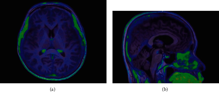
Methods: Twenty-six cognitively normal (CN) subjects, 7 patients with AD, and 36 patients with MCI were enrolled. A dynamic positron emission tomography (PET) scan was performed 30-75 min postinjection. PET and T1-weighted magnetic resonance imaging scans were coregistered. The standardized uptake value ratio (SUVr) was used for semiquantitative analysis. The P-Mod software was applied to create volumes of interest. The ANOVA and post hoc Tukey HSD were used for statistical analysis.
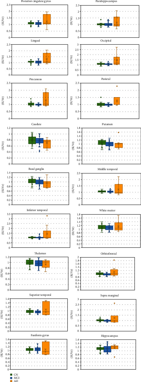
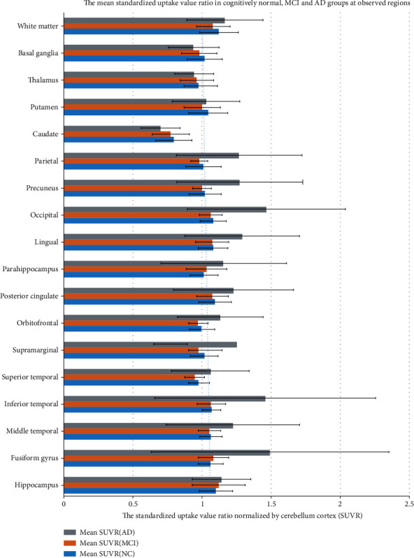
Results: In the AD group, the occipital lobe had a significantly higher mean SUVr (1.46 ± 0.57) than in the CN and MCI groups. Compared with the CN group, the AD group showed significantly higher mean SUVr in the fusiform gyrus (1.06 ± 0.09 vs. 1.49 ± 0.86), inferior temporal (1.07 ± 0.07 vs. 1.46 ± 0.08), parietal lobe, lingual gyrus, and precuneus regions. Similarly, the AD group demonstrated a higher mean SUVr than the MCI group in the precuneus, lingual, inferior temporal, fusiform, supramarginal, orbitofrontal, and superior temporal regions. The remaining observed regions, including the striatum, basal ganglia, thalamus, and white matter, showed a low SUVr across all groups with no statistically significant differences.
Conclusion: A significantly higher mean SUVr of [18F]PI-2620 was observed in the AD group; a significant area of the brain in the AD group demonstrated tau protein deposit in concordance with Braak Stages III-V, providing useful information to differentiate AD from CN and MCI. Moreover, the low SUVr in the deep striatum and thalamus could be useful for excluding primary tauopathies.
Molecular ImagingBiochemistry, Genetics and Molecular Biology-Biotechnology
自引率
3.60%
发文量
21
期刊介绍:
Molecular Imaging is a peer-reviewed, open access journal highlighting the breadth of molecular imaging research from basic science to preclinical studies to human applications. This serves both the scientific and clinical communities by disseminating novel results and concepts relevant to the biological study of normal and disease processes in both basic and translational studies ranging from mice to humans.
 求助内容:
求助内容: 应助结果提醒方式:


