Montserrat Diaz-Abad, Kathryn S Robinett, Anayansi Lasso-Pirot, Teklu B Legesse, Mariam Khambaty
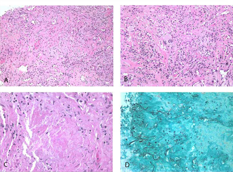
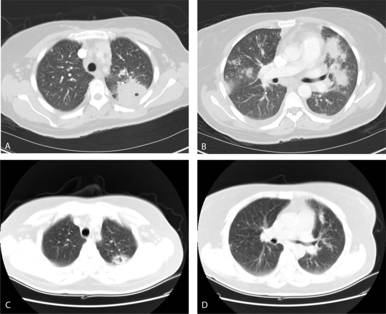
{"title":"Granulomatous <i>Pneumocystis jiroveci</i> Pneumonia in an HIV-Positive Patient on Antiretroviral Therapy: A Diagnostic Challenge.","authors":"Montserrat Diaz-Abad, Kathryn S Robinett, Anayansi Lasso-Pirot, Teklu B Legesse, Mariam Khambaty","doi":"10.2174/1874306402115010019","DOIUrl":null,"url":null,"abstract":"<p><p>Human Immunodeficiency Virus (HIV)-related Opportunistic Infections (OI), including <i>Pneumocystis jiroveci</i> pneumonia (PCP), have become much less commonplace with anti-retroviral therapy (ART). Despite this, OIs are still common and it is important to remain vigilant for their presence and be aware of how ART and OI chemoprophylaxis may lead to atypical disease presentations. We present the case of a 51-year-old woman with HIV and CD4+ T helper lymphocytes cell count > 200 cells/ul on both ART and trimethoprim/sulfamethoxazole prophylaxis who presented with cavitating lung masses, mediastinal lymphadenopathy and pleural effusions. Negative bronchoalveolar lavage (BAL) and transbronchial biopsy (TBBx) prompted a second diagnostic procedure with a transthoracic core needle biopsy; the final diagnosis was granulomatous PCP. This case showcases a very rare presentation of PCP, with both large cavitating lung masses on imaging and granulomatous reaction on pathology, as well as the challenge of a potentially missed diagnosis with negative BAL and TBBx requiring transthoracic core needle biopsy for a final diagnosis.</p>","PeriodicalId":39127,"journal":{"name":"Open Respiratory Medicine Journal","volume":"15 ","pages":"19-22"},"PeriodicalIF":0.0000,"publicationDate":"2021-06-18","publicationTypes":"Journal Article","fieldsOfStudy":null,"isOpenAccess":false,"openAccessPdf":"https://www.ncbi.nlm.nih.gov/pmc/articles/PMC8227459/pdf/","citationCount":"2","resultStr":null,"platform":"Semanticscholar","paperid":null,"PeriodicalName":"Open Respiratory Medicine Journal","FirstCategoryId":"1085","ListUrlMain":"https://doi.org/10.2174/1874306402115010019","RegionNum":0,"RegionCategory":null,"ArticlePicture":[],"TitleCN":null,"AbstractTextCN":null,"PMCID":null,"EPubDate":"2021/1/1 0:00:00","PubModel":"eCollection","JCR":"Q3","JCRName":"Medicine","Score":null,"Total":0}
引用次数: 2
Abstract
Human Immunodeficiency Virus (HIV)-related Opportunistic Infections (OI), including Pneumocystis jiroveci pneumonia (PCP), have become much less commonplace with anti-retroviral therapy (ART). Despite this, OIs are still common and it is important to remain vigilant for their presence and be aware of how ART and OI chemoprophylaxis may lead to atypical disease presentations. We present the case of a 51-year-old woman with HIV and CD4+ T helper lymphocytes cell count > 200 cells/ul on both ART and trimethoprim/sulfamethoxazole prophylaxis who presented with cavitating lung masses, mediastinal lymphadenopathy and pleural effusions. Negative bronchoalveolar lavage (BAL) and transbronchial biopsy (TBBx) prompted a second diagnostic procedure with a transthoracic core needle biopsy; the final diagnosis was granulomatous PCP. This case showcases a very rare presentation of PCP, with both large cavitating lung masses on imaging and granulomatous reaction on pathology, as well as the challenge of a potentially missed diagnosis with negative BAL and TBBx requiring transthoracic core needle biopsy for a final diagnosis.
期刊介绍:
The Open Respiratory Medicine Journal is an Open Access online journal, which publishes research articles, reviews/mini-reviews, letters and guest edited single topic issues in all important areas of experimental and clinical research in respiratory medicine. Topics covered include: -COPD- Occupational disorders, and the role of allergens and pollutants- Asthma- Allergy- Non-invasive ventilation- Therapeutic intervention- Lung cancer- Lung infections respiratory diseases- Therapeutic interventions- Adult and paediatric medicine- Cell biology. The Open Respiratory Medicine Journal, a peer reviewed journal, is an important and reliable source of current information on important recent developments in the field. The emphasis will be on publishing quality articles rapidly and making them freely available worldwide.

 求助内容:
求助内容: 应助结果提醒方式:


