Bart Witjes, Sylvain Baillet, Mathieu Roy, Robert Oostenveld, Frank J P M Huygen, Cecile C de Vos
{"title":"Magnetoencephalography reveals increased slow-to-fast alpha power ratios in patients with chronic pain.","authors":"Bart Witjes, Sylvain Baillet, Mathieu Roy, Robert Oostenveld, Frank J P M Huygen, Cecile C de Vos","doi":"10.1097/PR9.0000000000000928","DOIUrl":null,"url":null,"abstract":"<p><strong>Introduction: </strong>Objective disease markers are a key for diagnosis and personalized interventions. In chronic pain, such markers are still not available, and therapy relies on individual patients' reports. However, several pain studies have reported group-based differences in functional magnetic resonance imaging, electroencephalography, and magnetoencephalography (MEG).</p><p><strong>Objectives: </strong>We aimed to explore spectral differences in resting-state MEG brain signals between patients with chronic pain and pain-free controls and to characterize the cortical and subcortical regions involved.</p><p><strong>Methods: </strong>We estimated power spectral density over 5 minutes of resting-state MEG recordings in patients with chronic pain and controls and derived 7 spectral features at the sensor and source levels: alpha peak frequency, alpha power ratio (power 7-9 Hz divided by power 9-11 Hz), and average power in theta, alpha, beta, low-gamma, and high-gamma bands. We performed nonparametric permutation <i>t</i> tests (false discovery rate corrected) to assess between-group differences in these 7 spectral features.</p><p><strong>Results: </strong>Twenty-one patients with chronic pain and 25 controls were included. No significant group differences were found in alpha peak frequency or average power in any frequency band. The alpha power ratio was significantly higher (<i>P</i> < 0.05) in patients with chronic pain at both the sensor and brain source levels. The brain regions showing significantly higher ratios included the occipital, parietal, temporal and frontal lobe areas, insular and cingulate cortex, and right thalamus.</p><p><strong>Conclusion: </strong>The alpha power ratio is a simple, promising signal marker of chronic pain, affecting an expansive range of cortical and subcortical regions, including known pain-processing areas.</p>","PeriodicalId":3,"journal":{"name":"ACS Applied Electronic Materials","volume":" ","pages":"e928"},"PeriodicalIF":4.7000,"publicationDate":"2021-06-03","publicationTypes":"Journal Article","fieldsOfStudy":null,"isOpenAccess":false,"openAccessPdf":"https://www.ncbi.nlm.nih.gov/pmc/articles/PMC8177875/pdf/","citationCount":"9","resultStr":null,"platform":"Semanticscholar","paperid":null,"PeriodicalName":"ACS Applied Electronic Materials","FirstCategoryId":"1085","ListUrlMain":"https://doi.org/10.1097/PR9.0000000000000928","RegionNum":3,"RegionCategory":"材料科学","ArticlePicture":[],"TitleCN":null,"AbstractTextCN":null,"PMCID":null,"EPubDate":"2021/7/1 0:00:00","PubModel":"eCollection","JCR":"Q1","JCRName":"ENGINEERING, ELECTRICAL & ELECTRONIC","Score":null,"Total":0}
引用次数: 9
Abstract
Introduction: Objective disease markers are a key for diagnosis and personalized interventions. In chronic pain, such markers are still not available, and therapy relies on individual patients' reports. However, several pain studies have reported group-based differences in functional magnetic resonance imaging, electroencephalography, and magnetoencephalography (MEG).
Objectives: We aimed to explore spectral differences in resting-state MEG brain signals between patients with chronic pain and pain-free controls and to characterize the cortical and subcortical regions involved.
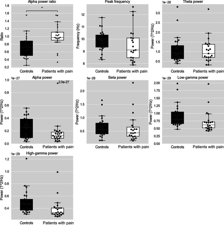
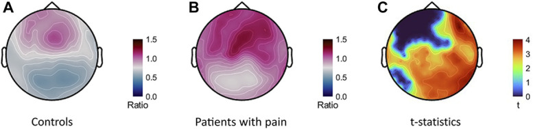
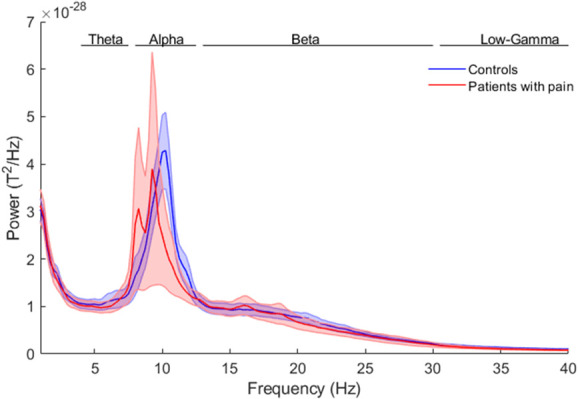
Methods: We estimated power spectral density over 5 minutes of resting-state MEG recordings in patients with chronic pain and controls and derived 7 spectral features at the sensor and source levels: alpha peak frequency, alpha power ratio (power 7-9 Hz divided by power 9-11 Hz), and average power in theta, alpha, beta, low-gamma, and high-gamma bands. We performed nonparametric permutation t tests (false discovery rate corrected) to assess between-group differences in these 7 spectral features.
Results: Twenty-one patients with chronic pain and 25 controls were included. No significant group differences were found in alpha peak frequency or average power in any frequency band. The alpha power ratio was significantly higher (P < 0.05) in patients with chronic pain at both the sensor and brain source levels. The brain regions showing significantly higher ratios included the occipital, parietal, temporal and frontal lobe areas, insular and cingulate cortex, and right thalamus.
Conclusion: The alpha power ratio is a simple, promising signal marker of chronic pain, affecting an expansive range of cortical and subcortical regions, including known pain-processing areas.
期刊介绍:
ACS Applied Electronic Materials is an interdisciplinary journal publishing original research covering all aspects of electronic materials. The journal is devoted to reports of new and original experimental and theoretical research of an applied nature that integrate knowledge in the areas of materials science, engineering, optics, physics, and chemistry into important applications of electronic materials. Sample research topics that span the journal's scope are inorganic, organic, ionic and polymeric materials with properties that include conducting, semiconducting, superconducting, insulating, dielectric, magnetic, optoelectronic, piezoelectric, ferroelectric and thermoelectric.
Indexed/Abstracted:
Web of Science SCIE
Scopus
CAS
INSPEC
Portico
 求助内容:
求助内容: 应助结果提醒方式:


