{"title":"Transcription factor signal transducer and activator of transcription 6 (STAT6) is an inhibitory factor for adult myogenesis.","authors":"Mitsutoshi Kurosaka, Yuji Ogura, Shuichi Sato, Kazuhisa Kohda, Toshiya Funabashi","doi":"10.1186/s13395-021-00271-8","DOIUrl":null,"url":null,"abstract":"<p><strong>Background: </strong>The signal transducer and activator of transcription 6 (STAT6) transcription factor plays a vitally important role in immune cells, where it is activated mainly by interleukin-4 (IL-4). Because IL-4 is an essential cytokine for myotube formation, STAT6 might also be involved in myogenesis as part of IL-4 signaling. This study was conducted to elucidate the role of STAT6 in adult myogenesis in vitro and in vivo.</p><p><strong>Methods: </strong>Myoblasts were isolated from male mice and were differentiated on a culture dish to evaluate the change in STAT6 during myotube formation. Then, the effects of STAT6 overexpression and inhibition on proliferation, differentiation, and fusion in those cells were studied. Additionally, to elucidate the myogenic role of STAT6 in vivo, muscle regeneration after injury was evaluated in STAT6 knockout mice.</p><p><strong>Results: </strong>IL-4 can increase STAT6 phosphorylation, but STAT6 phosphorylation decreased during myotube formation in culture. STAT6 overexpression decreased, but STAT6 knockdown increased the differentiation index and the fusion index. Results indicate that STAT6 inhibited myogenin protein expression. Results of in vivo experiments show that STAT6 knockout mice exhibited better regeneration than wild-type mice 5 days after cardiotoxin-induced injury. It is particularly interesting that results obtained using cells from STAT6 knockout mice suggest that this STAT6 inhibitory action for myogenesis was not mediated by IL-4 but might instead be associated with p38 mitogen-activated protein kinase phosphorylation. However, STAT6 was not involved in the proliferation of myogenic cells in vitro and in vivo.</p><p><strong>Conclusion: </strong>Results suggest that STAT6 functions as an inhibitor of adult myogenesis. Moreover, results suggest that the IL-4-STAT6 signaling axis is unlikely to be responsible for myotube formation.</p>","PeriodicalId":21747,"journal":{"name":"Skeletal Muscle","volume":null,"pages":null},"PeriodicalIF":5.3000,"publicationDate":"2021-05-29","publicationTypes":"Journal Article","fieldsOfStudy":null,"isOpenAccess":false,"openAccessPdf":"https://sci-hub-pdf.com/10.1186/s13395-021-00271-8","citationCount":"5","resultStr":null,"platform":"Semanticscholar","paperid":null,"PeriodicalName":"Skeletal Muscle","FirstCategoryId":"3","ListUrlMain":"https://doi.org/10.1186/s13395-021-00271-8","RegionNum":2,"RegionCategory":"医学","ArticlePicture":[],"TitleCN":null,"AbstractTextCN":null,"PMCID":null,"EPubDate":"","PubModel":"","JCR":"Q2","JCRName":"CELL BIOLOGY","Score":null,"Total":0}
引用次数: 5
Abstract
Background: The signal transducer and activator of transcription 6 (STAT6) transcription factor plays a vitally important role in immune cells, where it is activated mainly by interleukin-4 (IL-4). Because IL-4 is an essential cytokine for myotube formation, STAT6 might also be involved in myogenesis as part of IL-4 signaling. This study was conducted to elucidate the role of STAT6 in adult myogenesis in vitro and in vivo.
Methods: Myoblasts were isolated from male mice and were differentiated on a culture dish to evaluate the change in STAT6 during myotube formation. Then, the effects of STAT6 overexpression and inhibition on proliferation, differentiation, and fusion in those cells were studied. Additionally, to elucidate the myogenic role of STAT6 in vivo, muscle regeneration after injury was evaluated in STAT6 knockout mice.
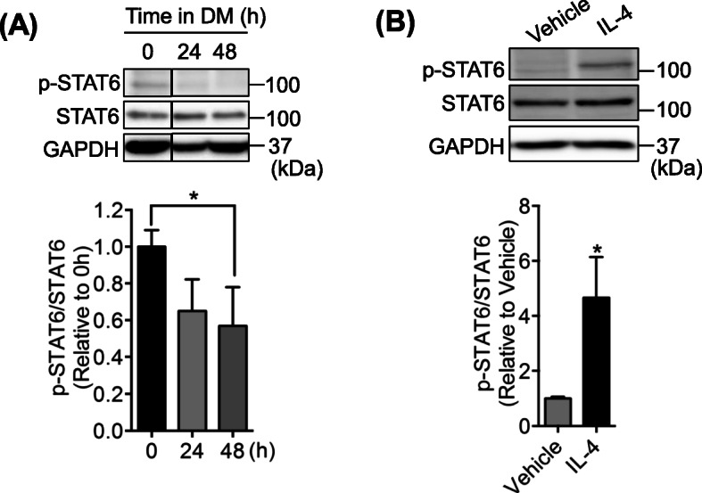
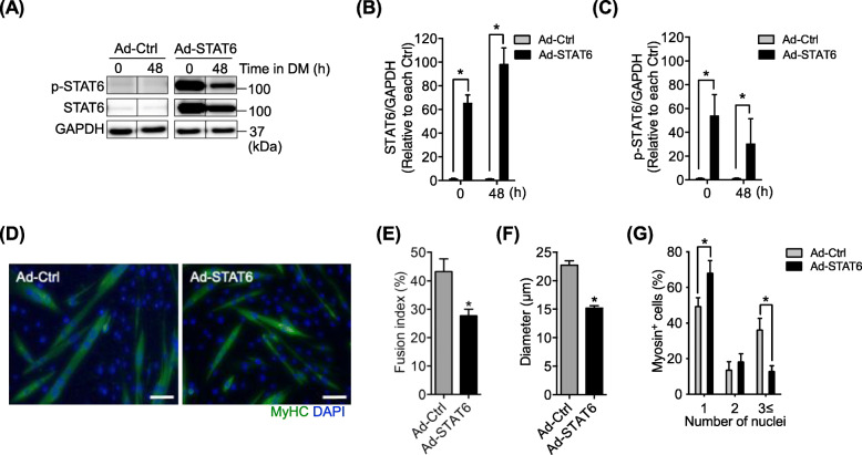
Results: IL-4 can increase STAT6 phosphorylation, but STAT6 phosphorylation decreased during myotube formation in culture. STAT6 overexpression decreased, but STAT6 knockdown increased the differentiation index and the fusion index. Results indicate that STAT6 inhibited myogenin protein expression. Results of in vivo experiments show that STAT6 knockout mice exhibited better regeneration than wild-type mice 5 days after cardiotoxin-induced injury. It is particularly interesting that results obtained using cells from STAT6 knockout mice suggest that this STAT6 inhibitory action for myogenesis was not mediated by IL-4 but might instead be associated with p38 mitogen-activated protein kinase phosphorylation. However, STAT6 was not involved in the proliferation of myogenic cells in vitro and in vivo.
Conclusion: Results suggest that STAT6 functions as an inhibitor of adult myogenesis. Moreover, results suggest that the IL-4-STAT6 signaling axis is unlikely to be responsible for myotube formation.
背景:转录因子STAT6 (signal transducer and activator of transcription factor)在免疫细胞中起着至关重要的作用,其主要被白细胞介素-4 (interleukin-4, IL-4)激活。由于IL-4是肌管形成的必需细胞因子,STAT6也可能作为IL-4信号传导的一部分参与肌生成。本研究旨在阐明STAT6在体外和体内成人肌发生中的作用。方法:从雄性小鼠中分离成肌细胞,在培养皿中分化,观察STAT6在肌管形成过程中的变化。然后,研究STAT6过表达和抑制对这些细胞增殖、分化和融合的影响。此外,为了阐明STAT6在体内的肌生成作用,我们在STAT6敲除小鼠中评估了损伤后的肌肉再生。结果:IL-4能提高STAT6的磷酸化水平,但在肌管形成过程中,STAT6的磷酸化水平降低。STAT6过表达降低,而STAT6敲低使分化指数和融合指数升高。结果表明,STAT6抑制了肌原蛋白的表达。体内实验结果表明,STAT6基因敲除小鼠在心脏毒素诱导损伤后5天的再生能力优于野生型小鼠。特别有趣的是,使用STAT6敲除小鼠的细胞获得的结果表明,这种STAT6对肌生成的抑制作用不是由IL-4介导的,而是可能与p38丝裂原激活的蛋白激酶磷酸化有关。然而,STAT6在体内和体外均未参与成肌细胞的增殖。结论:STAT6具有抑制成人肌生成的作用。此外,结果表明IL-4-STAT6信号轴不太可能负责肌管的形成。
期刊介绍:
The only open access journal in its field, Skeletal Muscle publishes novel, cutting-edge research and technological advancements that investigate the molecular mechanisms underlying the biology of skeletal muscle. Reflecting the breadth of research in this area, the journal welcomes manuscripts about the development, metabolism, the regulation of mass and function, aging, degeneration, dystrophy and regeneration of skeletal muscle, with an emphasis on understanding adult skeletal muscle, its maintenance, and its interactions with non-muscle cell types and regulatory modulators.
Main areas of interest include:
-differentiation of skeletal muscle-
atrophy and hypertrophy of skeletal muscle-
aging of skeletal muscle-
regeneration and degeneration of skeletal muscle-
biology of satellite and satellite-like cells-
dystrophic degeneration of skeletal muscle-
energy and glucose homeostasis in skeletal muscle-
non-dystrophic genetic diseases of skeletal muscle, such as Spinal Muscular Atrophy and myopathies-
maintenance of neuromuscular junctions-
roles of ryanodine receptors and calcium signaling in skeletal muscle-
roles of nuclear receptors in skeletal muscle-
roles of GPCRs and GPCR signaling in skeletal muscle-
other relevant aspects of skeletal muscle biology.
In addition, articles on translational clinical studies that address molecular and cellular mechanisms of skeletal muscle will be published. Case reports are also encouraged for submission.
Skeletal Muscle reflects the breadth of research on skeletal muscle and bridges gaps between diverse areas of science for example cardiac cell biology and neurobiology, which share common features with respect to cell differentiation, excitatory membranes, cell-cell communication, and maintenance. Suitable articles are model and mechanism-driven, and apply statistical principles where appropriate; purely descriptive studies are of lesser interest.

 求助内容:
求助内容: 应助结果提醒方式:


