{"title":"Altered Immunohistochemical Expression Patterns of HLA Class I during the Clinical Course of Cervical Intraepithelial Neoplasia.","authors":"Okuto Koguchi, Haruna Nishimaki, Yoko Nakanishi, Hiroko Kobayashi, Sumie Ohni, Xiaoyan Tang, Yoshiaki Kusumi, Shinobu Masuda","doi":"10.1267/ahc.21-00010","DOIUrl":null,"url":null,"abstract":"<p><p>It is unclear how immunohistochemical expression patterns of HLA class I in the pre-malignant phase of cervical intraepithelial neoplasia (CIN) alter during the clinical follow-up period. The present study aimed to demonstrate the correlation between the immunohistochemical expression pattern of HLA class I and the CIN grade through repeated examinations during the clinical course. Expression patterns of HLA class I, p16INK4a, and PD-L1 were immunohistochemically examined using formalin-fixed paraffin-embedded (FFPE) sections of biopsy or conization samples that were obtained from 20 patients diagnosed with CIN. The mRNA expression levels of HLA class I were analyzed by real-time reverse transcription polymerase chain reaction using FFPE sections of 14 patients, who were examined metachronously during the follow-up period. HLA class I expression was limited to the lower part of the epithelial thickness (M1 pattern) in more than half of CIN1 cases, and was present throughout the epithelial thickness (M2 pattern) in one fourth of CIN1 and CIN2 cases approximately. Heterogeneous expression (H pattern) was detected in half of CIN2 and CIN3 cases and in the all of squamous cell carcinoma cases. Metachronous examinations revealed that these immunohistochemical patterns altered more frequently than the CIN grade. The rate of change of HLA class I mRNA expression level was higher in cases with a progressed immunohistochemical pattern compared to those with regressed immunohistochemical pattern. In conclusion, the immunohistochemical pattern of HLA class I expression is associated with the CIN grade, and it is alterable during the clinical course, especially in CIN2.</p>","PeriodicalId":6888,"journal":{"name":"Acta Histochemica Et Cytochemica","volume":"54 2","pages":"57-64"},"PeriodicalIF":1.8000,"publicationDate":"2021-04-28","publicationTypes":"Journal Article","fieldsOfStudy":null,"isOpenAccess":false,"openAccessPdf":"https://ftp.ncbi.nlm.nih.gov/pub/pmc/oa_pdf/55/40/ahc-054-57.PMC8116618.pdf","citationCount":"0","resultStr":null,"platform":"Semanticscholar","paperid":null,"PeriodicalName":"Acta Histochemica Et Cytochemica","FirstCategoryId":"99","ListUrlMain":"https://doi.org/10.1267/ahc.21-00010","RegionNum":4,"RegionCategory":"生物学","ArticlePicture":[],"TitleCN":null,"AbstractTextCN":null,"PMCID":null,"EPubDate":"2021/4/17 0:00:00","PubModel":"Epub","JCR":"Q4","JCRName":"CELL BIOLOGY","Score":null,"Total":0}
引用次数: 0
Abstract
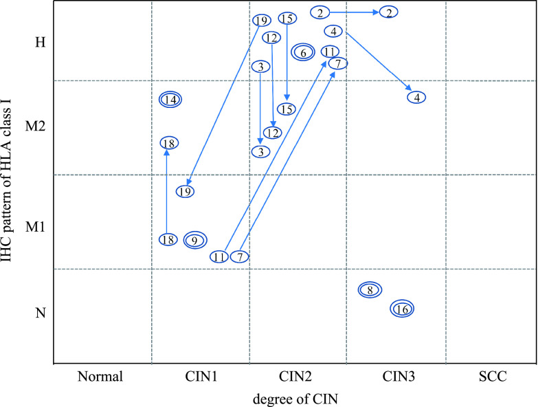
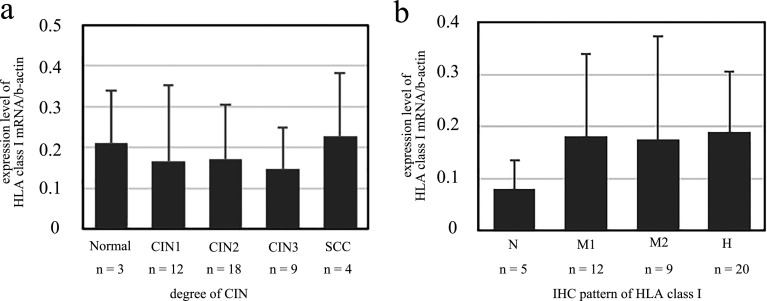
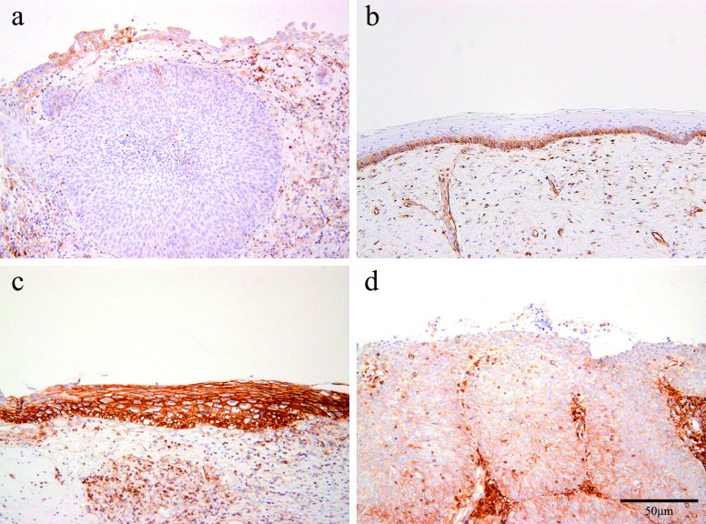
It is unclear how immunohistochemical expression patterns of HLA class I in the pre-malignant phase of cervical intraepithelial neoplasia (CIN) alter during the clinical follow-up period. The present study aimed to demonstrate the correlation between the immunohistochemical expression pattern of HLA class I and the CIN grade through repeated examinations during the clinical course. Expression patterns of HLA class I, p16INK4a, and PD-L1 were immunohistochemically examined using formalin-fixed paraffin-embedded (FFPE) sections of biopsy or conization samples that were obtained from 20 patients diagnosed with CIN. The mRNA expression levels of HLA class I were analyzed by real-time reverse transcription polymerase chain reaction using FFPE sections of 14 patients, who were examined metachronously during the follow-up period. HLA class I expression was limited to the lower part of the epithelial thickness (M1 pattern) in more than half of CIN1 cases, and was present throughout the epithelial thickness (M2 pattern) in one fourth of CIN1 and CIN2 cases approximately. Heterogeneous expression (H pattern) was detected in half of CIN2 and CIN3 cases and in the all of squamous cell carcinoma cases. Metachronous examinations revealed that these immunohistochemical patterns altered more frequently than the CIN grade. The rate of change of HLA class I mRNA expression level was higher in cases with a progressed immunohistochemical pattern compared to those with regressed immunohistochemical pattern. In conclusion, the immunohistochemical pattern of HLA class I expression is associated with the CIN grade, and it is alterable during the clinical course, especially in CIN2.
期刊介绍:
Acta Histochemica et Cytochemica is the official online journal of the Japan Society of Histochemistry and Cytochemistry. It is intended primarily for rapid publication of concise, original articles in the fields of histochemistry and cytochemistry. Manuscripts oriented towards methodological subjects that contain significant technical advances in these fields are also welcome. Manuscripts in English are accepted from investigators in any country, whether or not they are members of the Japan Society of Histochemistry and Cytochemistry. Manuscripts should be original work that has not been previously published and is not being considered for publication elsewhere, with the exception of abstracts. Manuscripts with essentially the same content as a paper that has been published or accepted, or is under consideration for publication, will not be considered. All submitted papers will be peer-reviewed by at least two referees selected by an appropriate Associate Editor. Acceptance is based on scientific significance, originality, and clarity. When required, a revised manuscript should be submitted within 3 months, otherwise it will be considered to be a new submission. The Editor-in-Chief will make all final decisions regarding acceptance.
 求助内容:
求助内容: 应助结果提醒方式:


